GLUTEOPLASTY · ANATOMICAL ATLAS

Gluteal Anatomy & Surface Contour

Understanding the structures that shape the buttock.

From deep muscular architecture to visible surface contour. A clinical reading of layers, landmarks and movement—before procedural planning.

Explore the anatomy
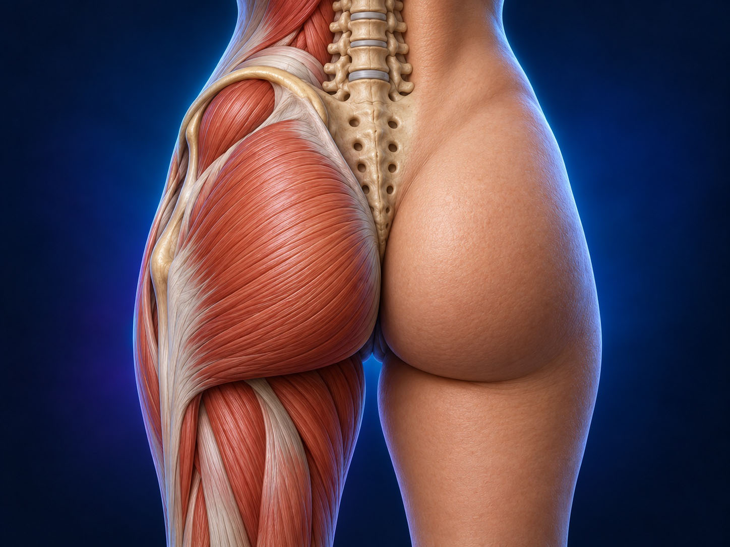
Planned illustration: Deep anatomy and visible surface contour ANATOMICAL ILLUSTRATION
Image placeholder
Deep anatomy and visible surface contour
01 · THE ESSENTIALS

Anatomy in 30 Seconds

Framework

Pelvis and sacrum establish the base. Muscles contribute bulk, force and stability.

Envelope

Fascia, septa, fat and skin translate deep architecture into visible shape.

Individual anatomy

Posture, contraction and tissue distribution change the reading. Surface landmarks cannot establish procedural safety.

02 · DEEP ARCHITECTURE

From Deep Anatomy to Surface Contour

Read the buttock as a layered anatomical system. The skeleton establishes the framework; muscles provide bulk and movement; fascia and fibrous septa connect tissues; subcutaneous fat and skin determine the visible envelope.

Clinical reading

A surface hollow does not identify a single deep structure. Assess several layers before attributing a contour to muscle, fat or tethering.

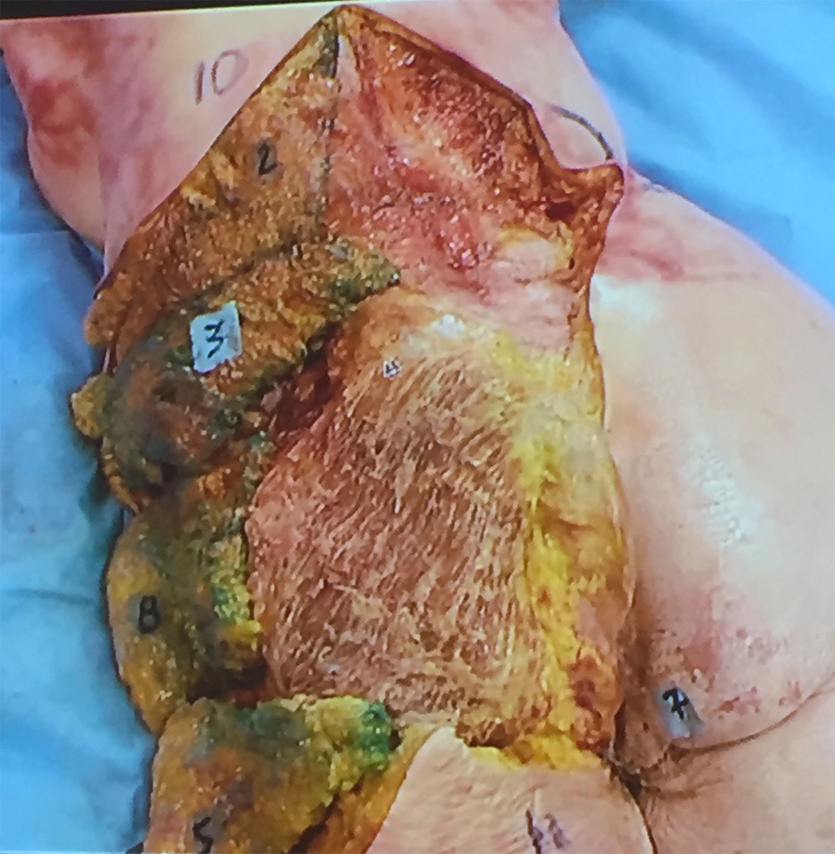
Cadaveric dissection showing gluteal subcutaneous fat and adjacent intact skin ANATOMICAL ILLUSTRATION
Image placeholder
Cadaveric view of gluteal subcutaneous fat and adjacent intact skin. Courtesy of Dr. Matt Stefanelli.
03 · DEEP ARCHITECTURE

Bony & Pelvic Framework

The iliac crest frames the upper region. The sacrum and coccyx form the central posterior axis. The posterior superior iliac spines (PSIS), greater trochanters and ischial tuberosities provide important orientation landmarks.

Clinical reading

Pelvic tilt and stance change apparent projection. Palpation and examination establish individual landmarks; a photograph alone cannot locate deep structures reliably.

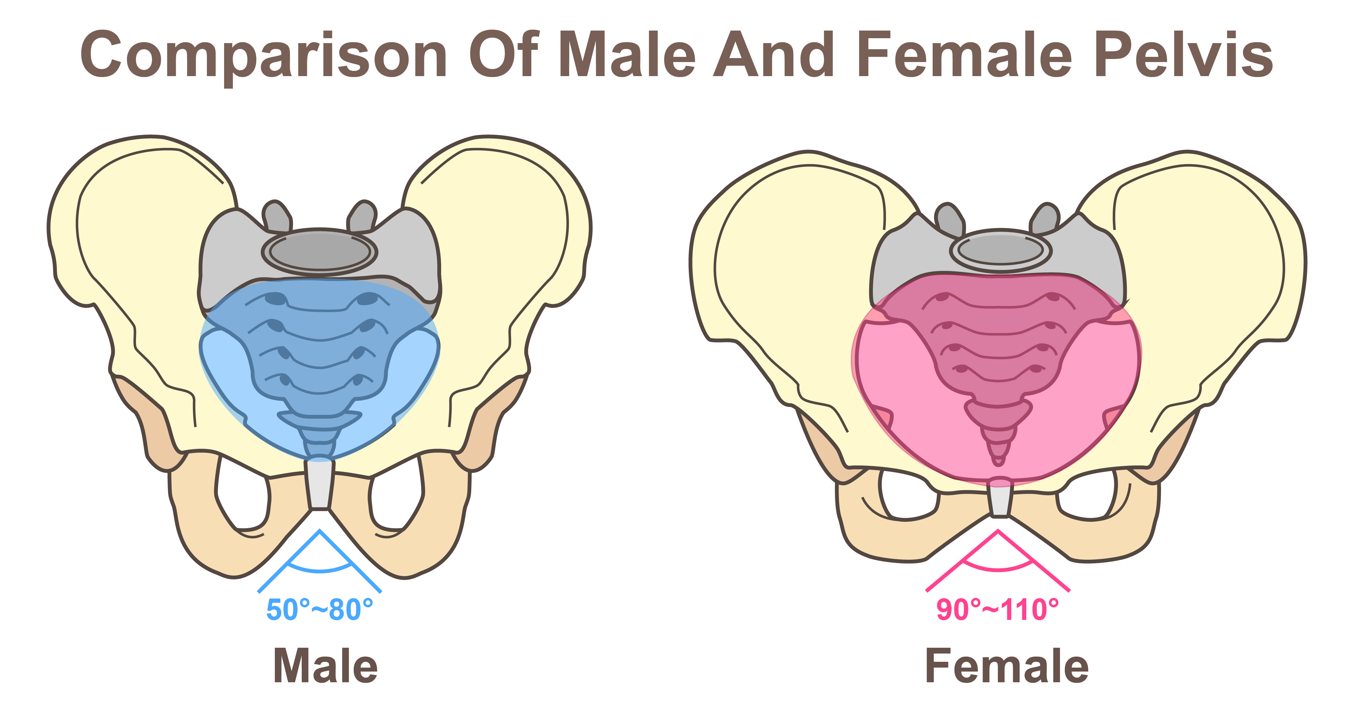
Anterior schematic comparison of male and female bony pelvis ANATOMICAL ILLUSTRATION
Image placeholder
Anterior schematic comparison of male and female pelvic structure. Individual anatomy varies; this diagram does not show the posterior surface landmarks.
04 · DEEP ARCHITECTURE

Gluteal Muscular Architecture

Gluteus maximus is the large superficial muscle. Gluteus medius lies mainly lateral and superior, partly covered by maximus; gluteus minimus lies deep to medius. The deeper short external rotators have distinct relationships to the hip and neurovascular structures.

Clinical reading

Maximus primarily extends and externally rotates the hip. Medius and minimus contribute to abduction and pelvic stability during single-leg support; actions vary with fibre position and joint angle.

Planned illustration: Gluteal Muscular Architecture ANATOMICAL ILLUSTRATION
Image placeholder
Gluteal Muscular Architecture
05 · DEEP ARCHITECTURE

Gluteus Maximus—The Main Contour Engine

The broad origin includes the posterior ilium, dorsal sacrum and coccyx, sacrotuberous ligament and associated fascia. Fibres pass inferolaterally to the iliotibial tract and gluteal tuberosity of the femur.

Clinical reading

Its size and architecture contribute strongly to posterior fullness. Visible projection also depends on fat distribution, pelvic orientation and the soft-tissue envelope; maximus alone does not determine shape.

Cadaveric dissection showing gluteus maximus and its visible muscle fibres ANATOMICAL ILLUSTRATION
Image placeholder
Cadaveric view of gluteus maximus and its visible fibres. Courtesy of Dr. Matt Stefanelli. This photograph does not show every origin and insertion.
06 · DEEP ARCHITECTURE

Fascia, Aponeuroses & Attachments

Gluteal fascia, the gluteal aponeurosis and continuity with the fascia lata support the region. Subcutaneous fibrous septa connect the skin and deeper tissues, influencing local mobility and transitions.

Clinical reading

Attachments are not interchangeable with wrinkles or muscle borders. A crease may reflect tethering, loading and envelope geometry rather than the edge of a muscle.

Planned illustration: Fascia, Aponeuroses & Attachments ANATOMICAL ILLUSTRATION
Image placeholder
Fascia, Aponeuroses & Attachments
07 · DEEP ARCHITECTURE

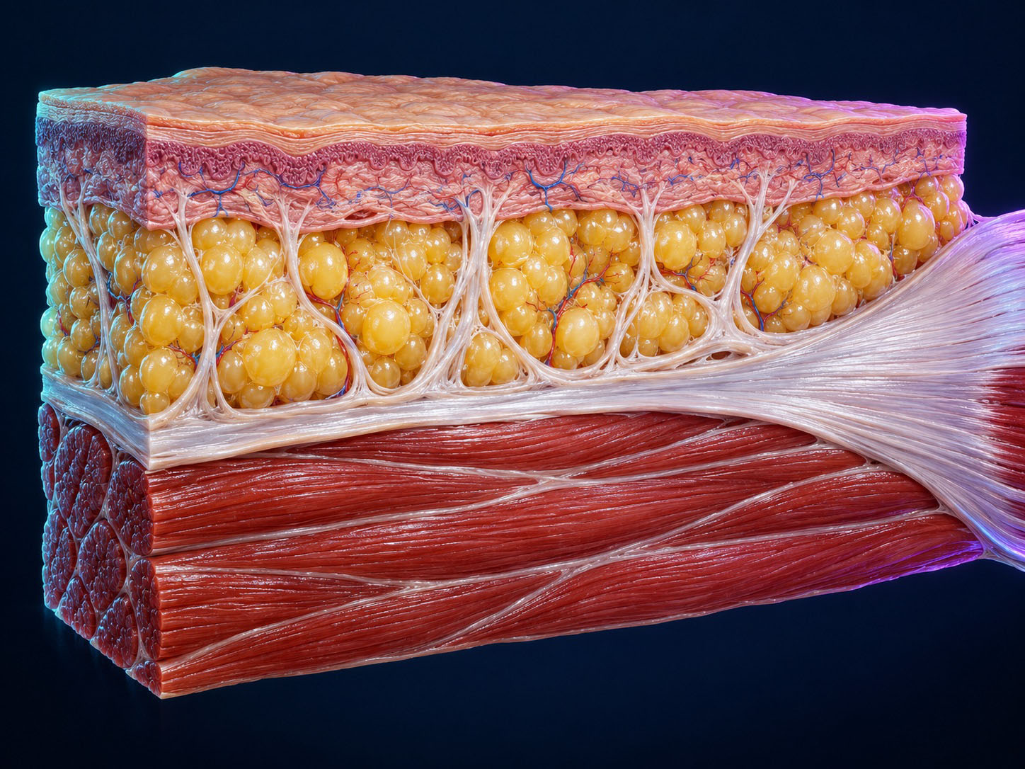
Subcutaneous Fat & Soft-Tissue Envelope

The subcutaneous layer contains adipose tissue organized by connective-tissue septa. Its thickness and distribution vary across the buttock and between individuals. Skin elasticity and tissue laxity further influence drape.

Clinical reading

Distinguish muscle bulk, adipose fullness, skin laxity and focal tethering. Similar surface contours can arise from different combinations of these tissues.

Cadaveric dissection showing gluteal subcutaneous fat and adjacent intact skin ANATOMICAL ILLUSTRATION
Image placeholder
Cadaveric view of gluteal subcutaneous fat and adjacent intact skin. Courtesy of Dr. Matt Stefanelli.
08 · SURFACE ATLAS

Surface Anatomical Landmarks

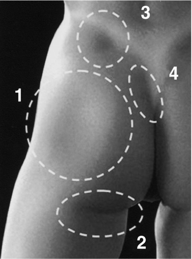
Examine the iliac crest, sacral midline, PSIS region, lateral trochanteric transition, intergluteal cleft and infragluteal folds together. Compare both sides using consistent stance and lighting.

Clinical reading

Surface landmarks orient examination. They are not a substitute for knowledge of depth, anatomical variation or procedure-specific safety assessment.

Photograph indicating lateral depression, infragluteal fold, supragluteal dimple and upper intergluteal crease ANATOMICAL ILLUSTRATION
Image placeholder
Selected surface features: 1 lateral depression; 2 infragluteal fold; 3 supragluteal dimple region; 4 upper intergluteal crease. Surface appearance does not establish the depth of underlying structures.
09 · SURFACE ATLAS

Presacral Triangle

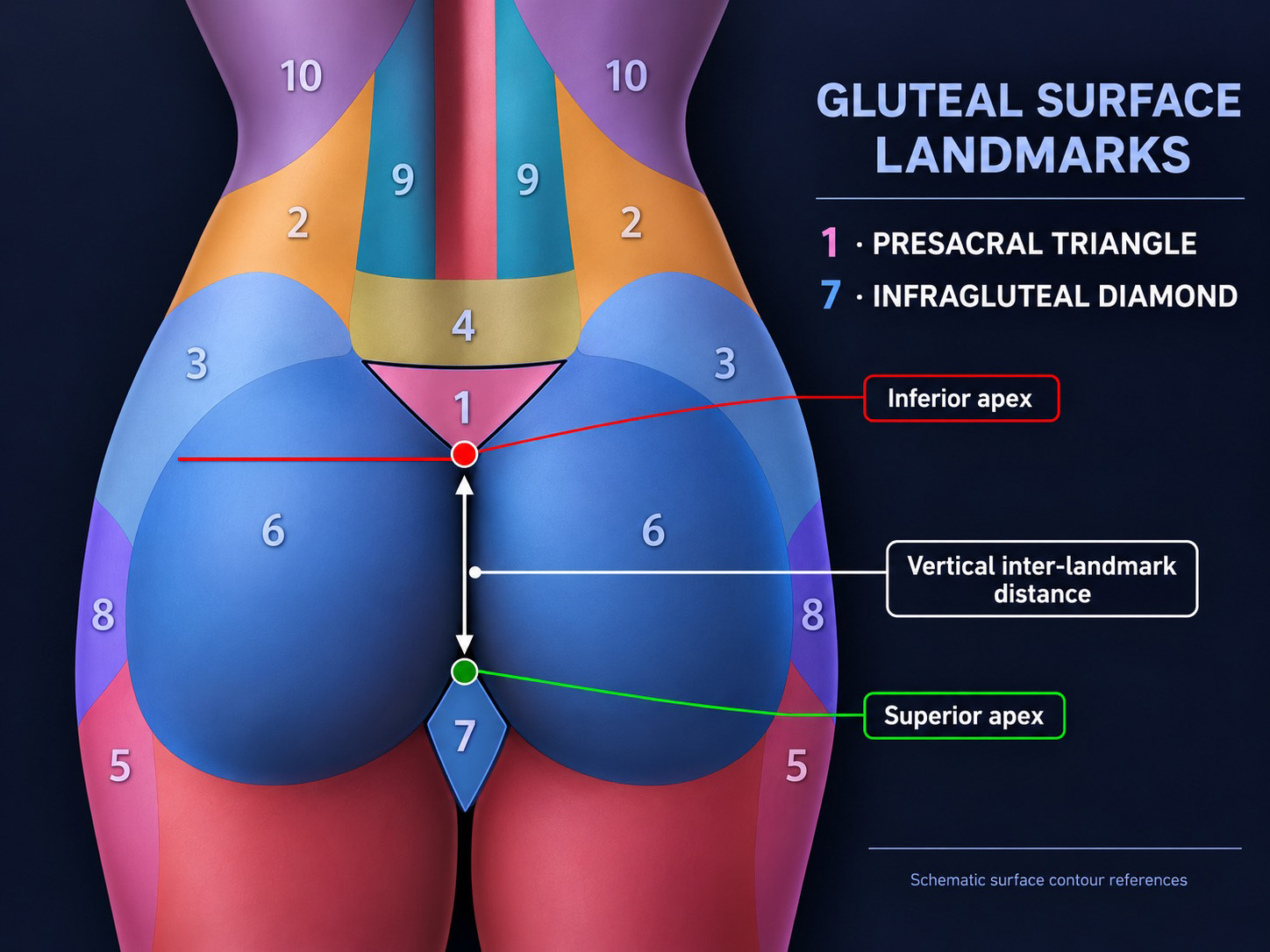
In this contour atlas, “presacral triangle” describes the posterior surface region over the sacrum between the upper buttocks. It is a descriptive visual reference, not a discrete muscle or a universally standardized anatomical compartment.

Clinical reading

Do not confuse this posterior surface descriptor with the internal presacral space anterior to the sacrum. Visibility varies with posture, adiposity and individual attachments.

Schematic posterior surface landmarks: presacral triangle, infragluteal diamond and vertical inter-landmark distance ANATOMICAL ILLUSTRATION
Image placeholder
Schematic surface landmarks: 1 presacral triangle; 7 infragluteal diamond. The vertical arrow connects the inferior apex of the triangle to the superior apex of the diamond.
10 · SURFACE ATLAS

Infragluteal Diamond

“Infragluteal diamond” is used here as a descriptive contour region at the medial buttock–upper-thigh transition. Its appearance relates to the lower cleft, paired folds and the position of the thighs.

Clinical reading

There are no universal anatomical borders or mandatory ideal proportions for this descriptor. Record observed transitions rather than inferring an isolated deep structure.

Schematic gluteal surface map showing the infragluteal diamond as region 7 and its superior apex in green ANATOMICAL ILLUSTRATION
Image placeholder
Region 7 identifies the infragluteal diamond; the green marker indicates its superior apex. This is a schematic surface contour reference, not a discrete deep anatomical compartment.
11 · SURFACE ATLAS

Lateral Depression / Hip Dip

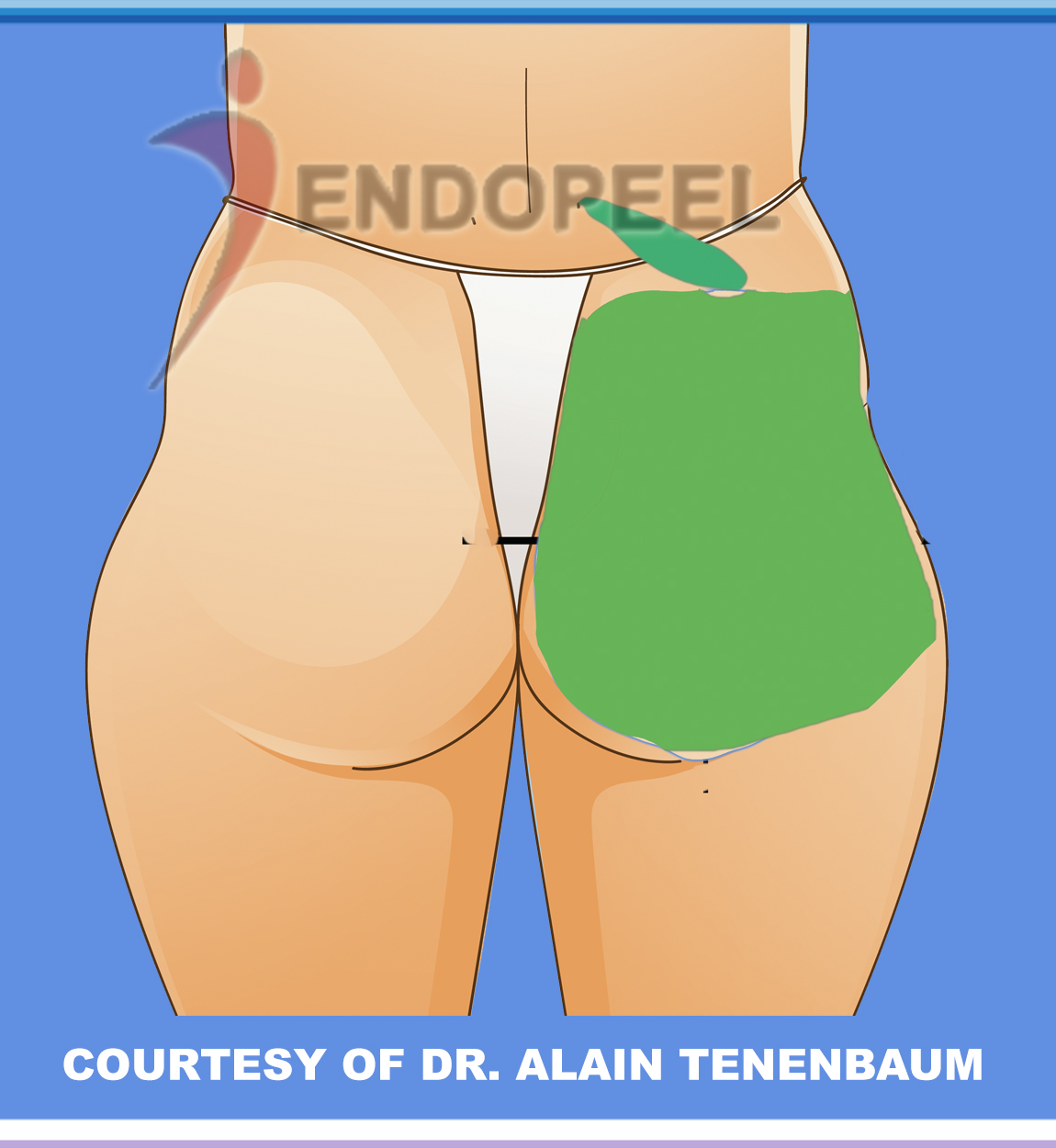
The lateral depression is a common variation at the transition between the pelvic region and greater trochanter. Pelvic geometry, muscle contours, fascial attachments and regional fat distribution all contribute to its appearance.

Clinical reading

A hip dip is not inherently a defect or disease. Its depth cannot be explained by a single insertion, and anatomy alone does not predict a treatment response.

Posterior contour schematic illustrating a lateral hip depression ANATOMICAL ILLUSTRATION
Image placeholder
Schematic lateral contour illustration. Courtesy of Dr. Alain Tenenbaum. The overlay does not identify individual deep structures or establish a treatment outcome.
12 · SURFACE ATLAS

Infra Gluteal Area

A region to examine—not just a crease to trace.

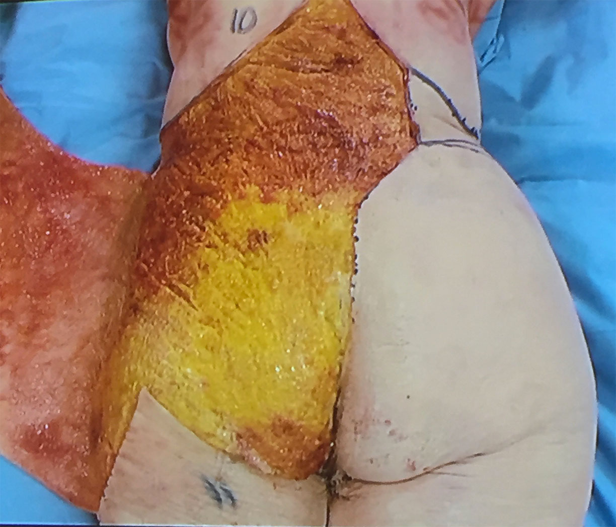
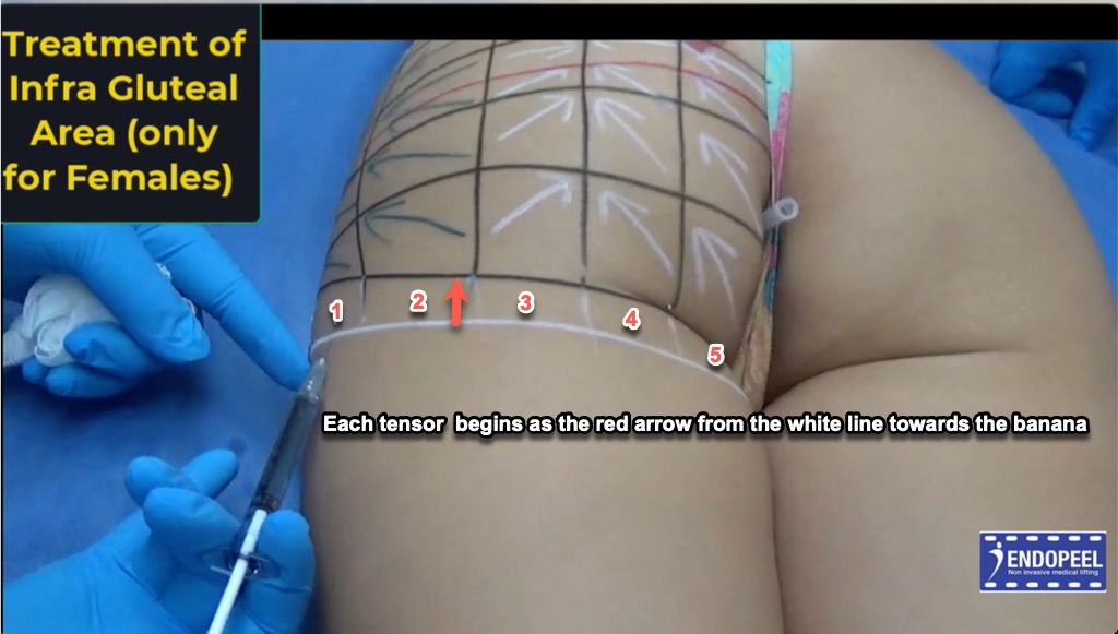
The infra gluteal area is the transition beneath the lower buttock into the proximal posterior thigh. In this atlas, it is assessed as a region with surface extent and tissue depth; the infragluteal fold is one visible landmark within this broader reading.

The superficial contour reflects skin, subcutaneous fat and fibrous septa, together with continuity of the fascial envelope toward the posterior thigh. Deeper relationships include the ischial tuberosity and proximal hamstring origins, with gluteus maximus overlying part of this transition. The appearance of the region does not reveal these structures or their depth directly.

Clinical reading

Assess subgluteal fullness, the buttock-to-thigh transition, skin laxity and symmetry separately from the crease. A localized fat roll below the fold, descent of buttock tissue and a pronounced fold are different observations; one does not establish the cause of another.

Compare relaxed standing with controlled contraction using matched posture and lighting. The sciatic nerve and posterior femoral cutaneous nerve and its branches are relevant deep relationships; the visible region is not a safe treatment zone.

Explore the Infra Gluteal Fold separately →

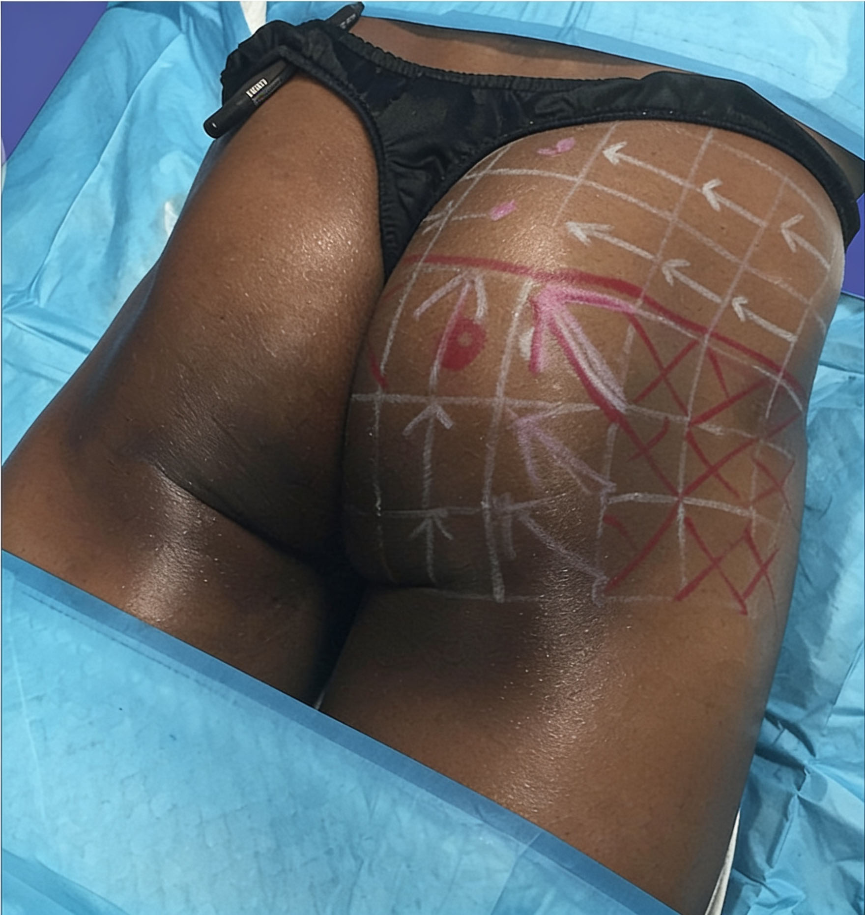
Historical clinical photograph showing the infra gluteal area below the infragluteal fold, with procedural markings ANATOMICAL ILLUSTRATION
Image placeholder
Historical clinical view locating the infra gluteal area below the fold. Procedural annotations belong to the source image; the visible contour does not establish tissue depth or a safe treatment zone.
13 · SURFACE ATLAS

Infragluteal Fold / Banana Fold

The infragluteal fold marks a buttock–posterior-thigh transition supported by connective-tissue anchoring and the arrangement of the soft-tissue envelope. It does not simply trace the inferior border of gluteus maximus.

Clinical reading

“Banana fold” is a colloquial term and may also refer to adjacent subgluteal fullness. Distinguish the crease from the fat roll below it and from true tissue descent.

Annotated surface photograph identifying the infragluteal fold in region 2 ANATOMICAL ILLUSTRATION
Image placeholder
Marker 2 indicates the infragluteal fold region. The crease is a surface landmark and should be distinguished from the broader infra gluteal area and adjacent subgluteal fullness.
14 · SURFACE ATLAS

Intergluteal Cleft

The intergluteal or natal cleft is the midline groove separating the buttocks over the sacrococcygeal region. Its visible depth and length depend on adjacent volume, skin attachments and position.

Clinical reading

Inspect the skin as well as the contour. Inflammation, drainage, pain or a suspected pilonidal condition calls for medical assessment before aesthetic planning.

Annotated surface photograph showing the intergluteal cleft, with its upper portion marked 4 ANATOMICAL ILLUSTRATION
Image placeholder
Marker 4 identifies the upper portion of the intergluteal cleft. The midline groove continues inferiorly between the buttocks; the annotation describes surface anatomy, not a treatment zone.
15 · SURFACE ATLAS

Supragluteal Dimples & V-Shaped Crease

Supragluteal dimples are often seen near the PSIS region, where local soft-tissue attachments influence the surface. A V-shaped crease may be visible at the upper cleft, with variable expression.

Clinical reading

Neither feature is obligatory. A fixed crease length, universal ratio or single causal muscle insertion should not be inferred from surface appearance.

Annotated surface photograph showing a supragluteal dimple at marker 3 and the upper cleft region at marker 4 ANATOMICAL ILLUSTRATION
Image placeholder
Marker 3 identifies the supragluteal dimple region; marker 4 indicates the upper intergluteal cleft. This photograph illustrates the dimple and adjacent surface landmarks, rather than a complete V-shaped crease.
16 · CLINICAL READING

Neurovascular Anatomy & Safety

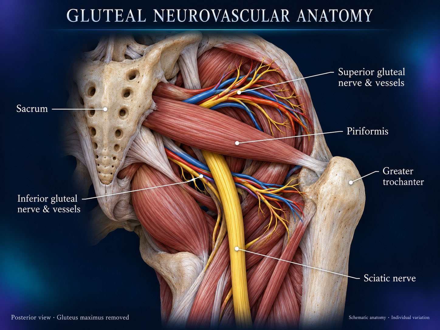
The superior gluteal neurovascular bundle typically leaves the pelvis above piriformis. The inferior gluteal bundle and sciatic nerve usually pass below it. Important vessels and nerves lie deep to gluteus maximus; branching and course vary.

Clinical reading

No visible triangle, quadrant or crease guarantees safety. Deep venous injury is a major concern in gluteal fat grafting; its safety guidance must not be generalized to other procedures. This atlas provides no injection sites, depths or dosing.

Planned illustration: Neurovascular Anatomy & Safety ANATOMICAL ILLUSTRATION
Image placeholder
Neurovascular Anatomy & Safety
17 · CLINICAL READING

Dynamic Anatomy—Relaxation vs Contraction

Muscle activation changes local firmness and contour; hip position and pelvic tilt alter what is seen. Compare relaxed standing with controlled contraction and, when relevant, movement or single-leg stance.

Clinical reading

Use matched views and lighting. A change in contour during contraction does not by itself demonstrate treatment efficacy or establish which tissue should be treated.

Paired male gluteal views at relaxation and contraction 30 minutes after unilateral myomodulation ANATOMICAL ILLUSTRATION
Image placeholder
Paired views at relaxation and contraction, 30 minutes after unilateral myomodulation; only the patient’s left side was treated. This individual observation illustrates dynamic contour assessment, not an untreated anatomical baseline or evidence of a durable result. Courtesy of Dr Alain Tenenbaum.
18 · CLINICAL READING

Male vs Female Gluteal Anatomy

Average sex-related differences in pelvic geometry, muscle mass and fat distribution may influence contour. Individual overlap is substantial, and age, training, body composition and posture also matter.

Clinical reading

Assess the person, not a stereotype. No universal male, female or ethnicity-based ideal can replace individual anatomy and patient-defined objectives.

Schematic comparison of male and female bony pelvic morphology ANATOMICAL ILLUSTRATION
Image placeholder
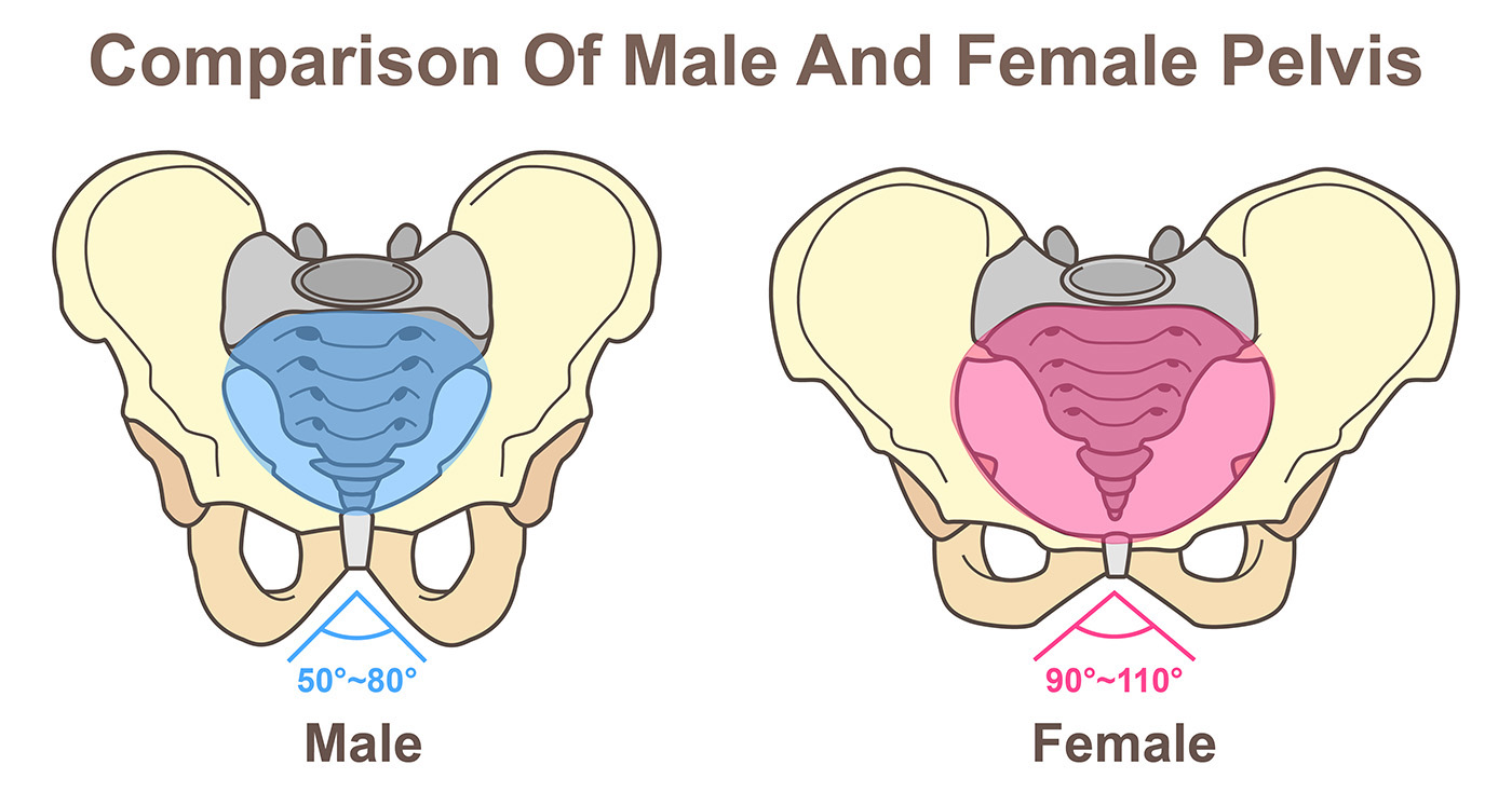
Schematic comparison of the bony pelvic framework, one contributor to gluteal contour. The illustrated angles are schematic references, not universal diagnostic limits; individual morphology varies. Muscle mass and fat distribution are not depicted.
19 · CLINICAL READING

From Anatomy to Myomodulation®

Anatomical assessment identifies the tissues contributing to a contour and their functional relationships. In this clinical vocabulary, Myomodulation® is the technique and concept; Endopeel® is a specific formulation used for myomodulation, not a generic product category.

Clinical reading

This page establishes anatomical context. Fundamental Basics addresses the 3D/isovolumetric deformation model; Step by Step addresses treatment execution. Anatomy is not evidence of efficacy or a guarantee of safety.

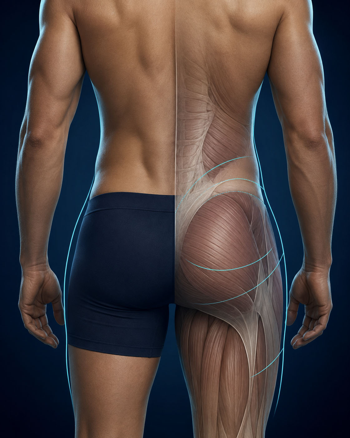
Conceptual illustration connecting male gluteal surface contour with underlying muscular architecture ANATOMICAL ILLUSTRATION
Image placeholder
Conceptual illustration linking visible contour with underlying muscular architecture. The turquoise lines are schematic graphic overlays, not validated anatomical boundaries or injection trajectories. This image provides anatomical context for Myomodulation®, without demonstrating a treatment result.
20 · CLINICAL READING

Clinical Anatomical Mapping

Document posture, pelvic orientation, palpable bony landmarks, symmetry, regional fullness, skin quality, creases and the response to contraction. Review previous surgery, scars, implants and relevant symptoms.

Clinical reading

Integrate clinical history, examination and appropriate imaging when indicated. Map what is observed, identify uncertainty and keep contour assessment separate from procedural execution.

Clinical photograph of individualized surface markings on the male lateral gluteal contour ANATOMICAL ILLUSTRATION
Image placeholder
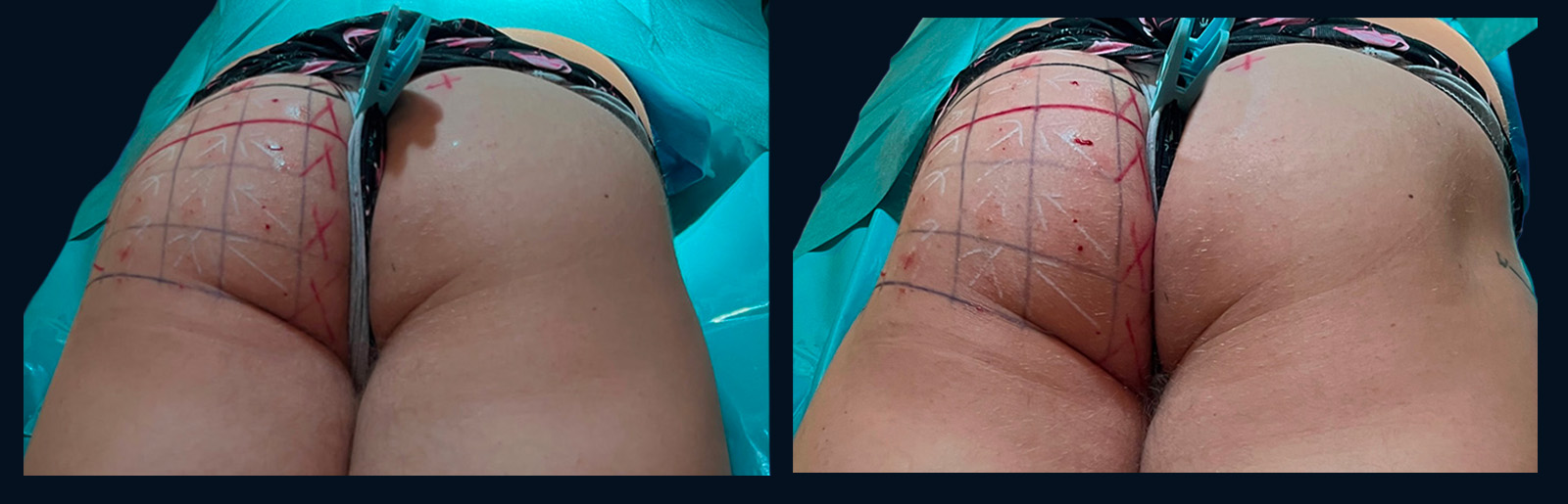
Clinical example of individualized surface marking on the lateral gluteal contour. Lines, arrows and crosses belong to this patient’s procedural planning; they are not universal anatomical boundaries, safe zones or injection instructions. Courtesy of Dr Alain Tenenbaum.
· QUESTIONS

Anatomy FAQ

How does the Infra Gluteal Area differ from the Infra Gluteal Fold?

The Infra Gluteal Area is the broader buttock–upper-posterior-thigh transition region. The Infra Gluteal Fold is a crease within that regional assessment. Fullness below the fold, tissue descent and the crease itself are separate findings.

What creates the visible buttock contour?

The combined effects of bone, muscle, fascia, subcutaneous fat, skin and posture. No single layer explains every contour.

Is the infragluteal fold the lower edge of gluteus maximus?

No. Connective-tissue anchoring and the soft-tissue envelope contribute to the fold; it does not simply reproduce the muscle border.

Are hip dips abnormal?

Usually they are a normal contour variation. Clinical assessment is needed for new, painful or otherwise concerning changes.

Are the presacral triangle and infragluteal diamond fixed anatomical compartments?

Here they are explicitly defined surface descriptors. They should not be treated as standardized deep compartments or safe treatment zones.

Can surface landmarks guarantee procedural safety?

No. Depth, variable neurovascular anatomy, procedure-specific guidance and clinician training remain essential.

How do Anatomy, Fundamental Basics and Step by Step differ?

Anatomy identifies structures creating the contour. Fundamental Basics explains the 3D/isovolumetric deformation model. Step by Step covers treatment execution.

Are Myomodulation® and Endopeel® interchangeable?

No. Myomodulation® names the technique/concept; Endopeel® names a specific formulation for myomodulation.

THE NEXT CHAPTER

From Understanding to Clinical Execution

Anatomy establishes which structures create the contour. Continue to the conceptual model or the dedicated procedural chapter.

Continue to Step by Step → Explore Fundamental Basics