Male gluteal contour viewed from behind

Male Gluteoplasty

Anatomy defines the starting point. Your goals define the direction.

Male gluteoplasty is not the reproduction of a standardized masculine shape. It is a personalized approach to gluteal contour, projection, symmetry and muscular balance, guided by anatomy, function and the individual objectives of each patient.

Anatomy-led Function-conscious Individually planned
Explore male gluteoplasty

Understanding the procedure

What Is Male Gluteoplasty?

Male gluteoplasty is a personalized approach to improving the contour, projection and balance of the male gluteal region.

Rather than reproducing a predetermined shape, treatment begins with an evaluation of the patient’s anatomy, muscular structure, tissue quality and individual objectives. The aim is to create a result that remains proportionate to the waist, pelvis, lower back and thighs.

Anatomical proportions and muscular structure

Projection, contour and gluteal symmetry

Individual goals and functional balance

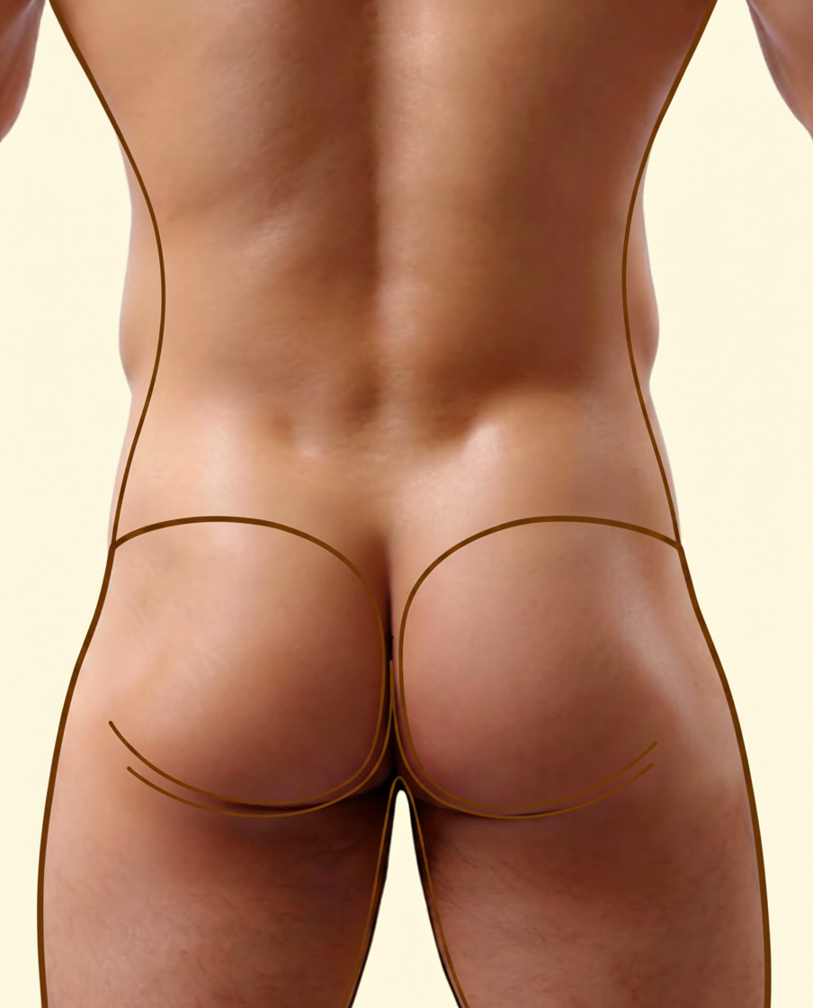
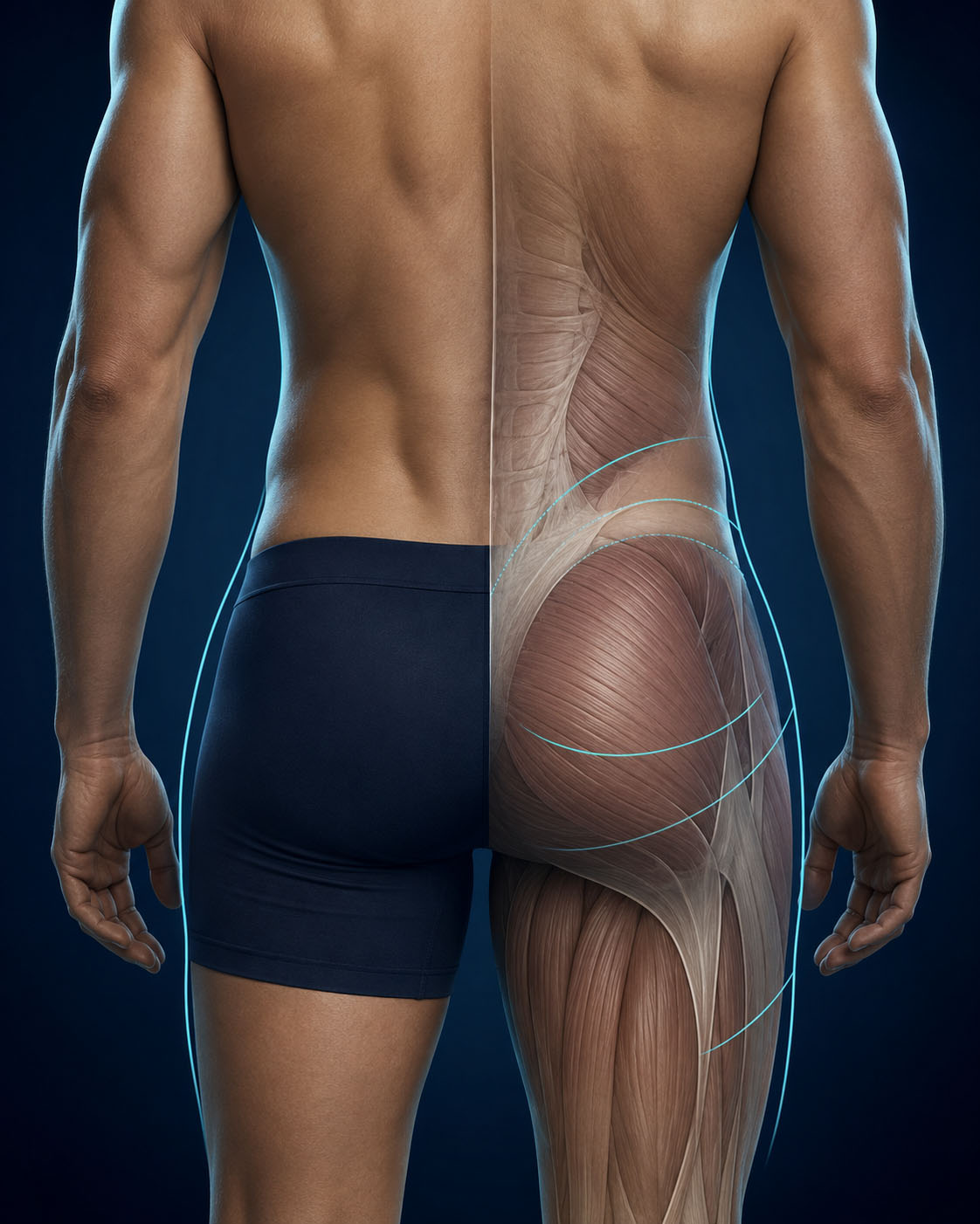
Male gluteal anatomy and muscular contour
Male gluteal anatomy varies in contour, projection and muscular balance.
Surface boundaries of the male gluteal region Upper boundary Lateral contour Infragluteal fold
Surface boundaries of the male gluteal region. Anatomical landmarks vary according to skeletal structure, muscular development and tissue distribution.

Form follows anatomy

Male Gluteal Anatomy

The male gluteal region is shaped by the relationship between the pelvis, muscular volume, fat distribution and the surrounding contours of the lower back and thighs.

Understanding these anatomical relationships is essential when planning male gluteoplasty.
Projection should not be confused with volume augmentation. Adding mass can distort masculine proportions and increase the downward load on the buttocks.
The objective is to improve projection without making the gluteal region heavier or more prone to descent. This is the principle of isovolumetric myomodulation..

Upper Boundary

The transition from the lower back to the upper gluteal region influences the apparent height and definition of the buttocks.


Lateral Contour

Pelvic width, muscular development and surrounding tissues shape the lateral relationship between the waist, hips and thighs.

Infragluteal Fold

The natural fold beneath each buttock defines the lower boundary and must be evaluated independently on both sides.

These surface landmarks provide a general anatomical reference. Individual anatomy must always be assessed during a clinical examination.

Different anatomies. Different objectives.

Why Men Consider Gluteoplasty

Men may consider gluteoplasty for different anatomical and personal reasons. The objective is not indiscriminate enlargement, but a precise response to projection, support, proportion, symmetry and contour.
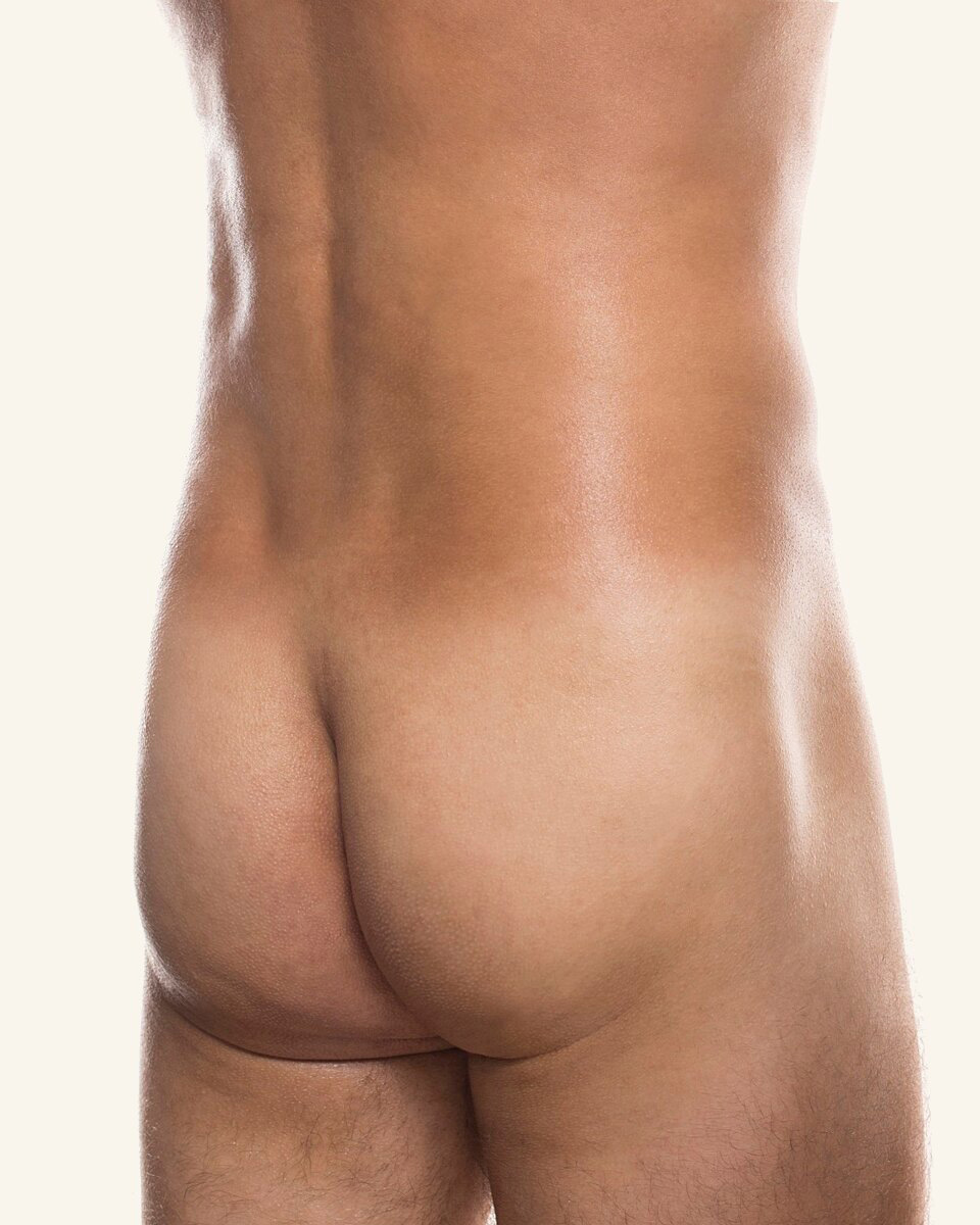
Oblique posterior view illustrating limited male gluteal projection

Projection

Insufficient Posterior Projection

A flat posterior profile may reflect pelvic structure, muscular morphology or individual tissue distribution.
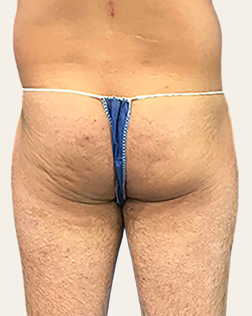
Posterior view illustrating loss of gluteal support in a male patient

Support & tissue position

Loss of Gluteal Support

Tissue laxity or inferior displacement may reduce firmness and alter the transition between the lower back, buttocks and upper thighs.
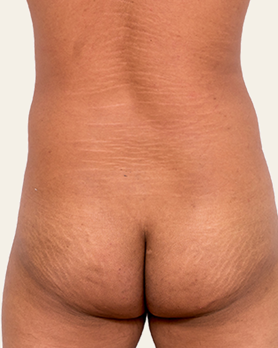
Posterior view illustrating the relationship between torso width and gluteal contour

Body proportions

Torso–Gluteal Disproportion

The relationship between torso width, waist structure and gluteal contour may create an imbalance in the posterior silhouette.
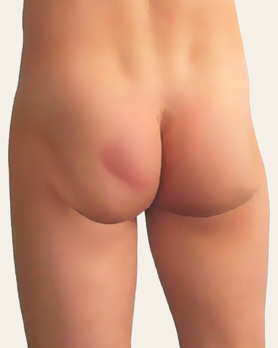
Posterior view illustrating marked asymmetry of the male gluteal contour

Balance & contour

Asymmetry or Contour Irregularities

Differences in volume, height or contour between the two sides may affect the overall balance and harmony of the gluteal region.
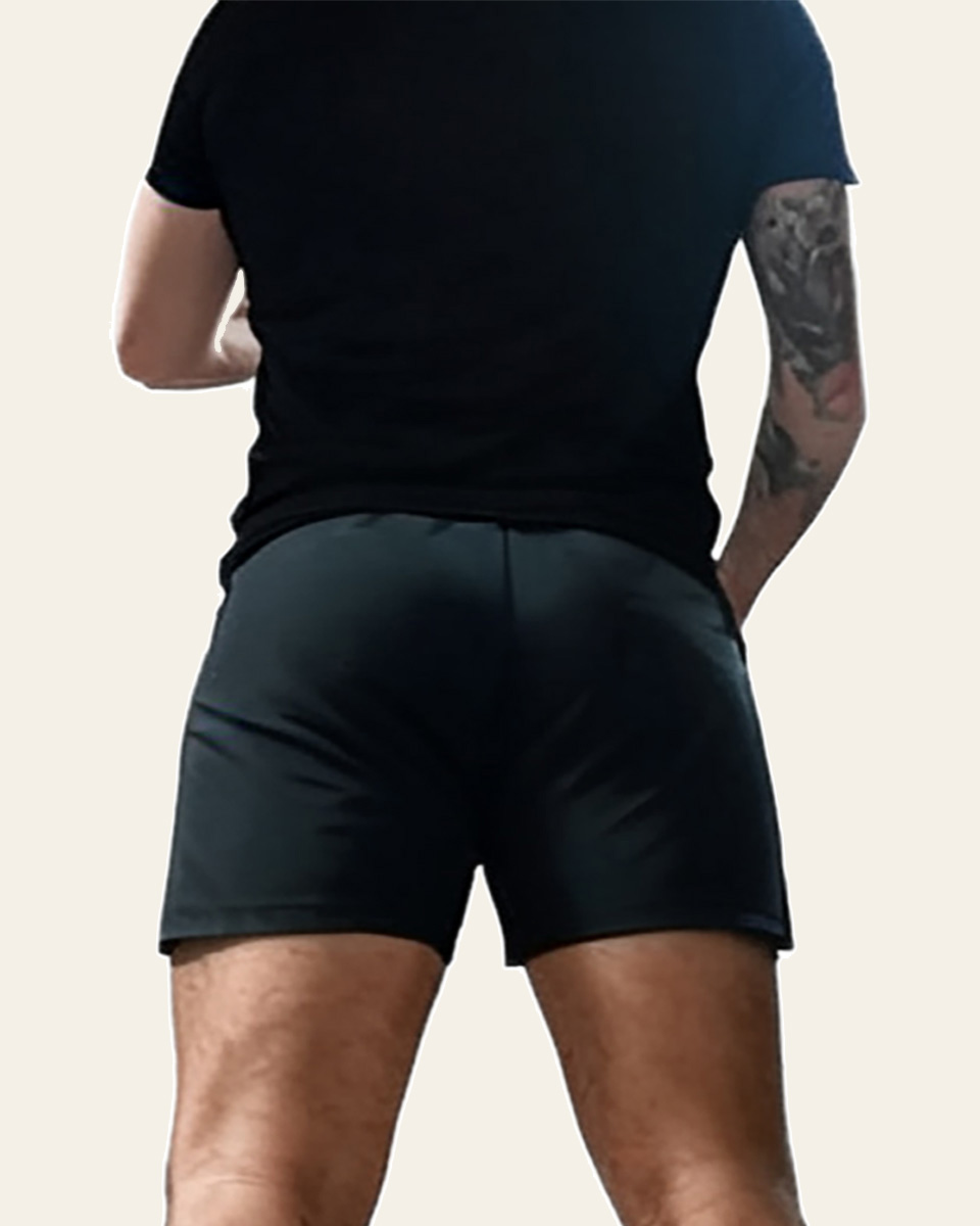
Athletic male posterior view illustrating structural limitations in gluteal projection

Anatomy & training

Structural Limitations Despite Training

Even with consistent training, skeletal structure, muscle insertions and fat distribution may limit achievable gluteal projection and definition.
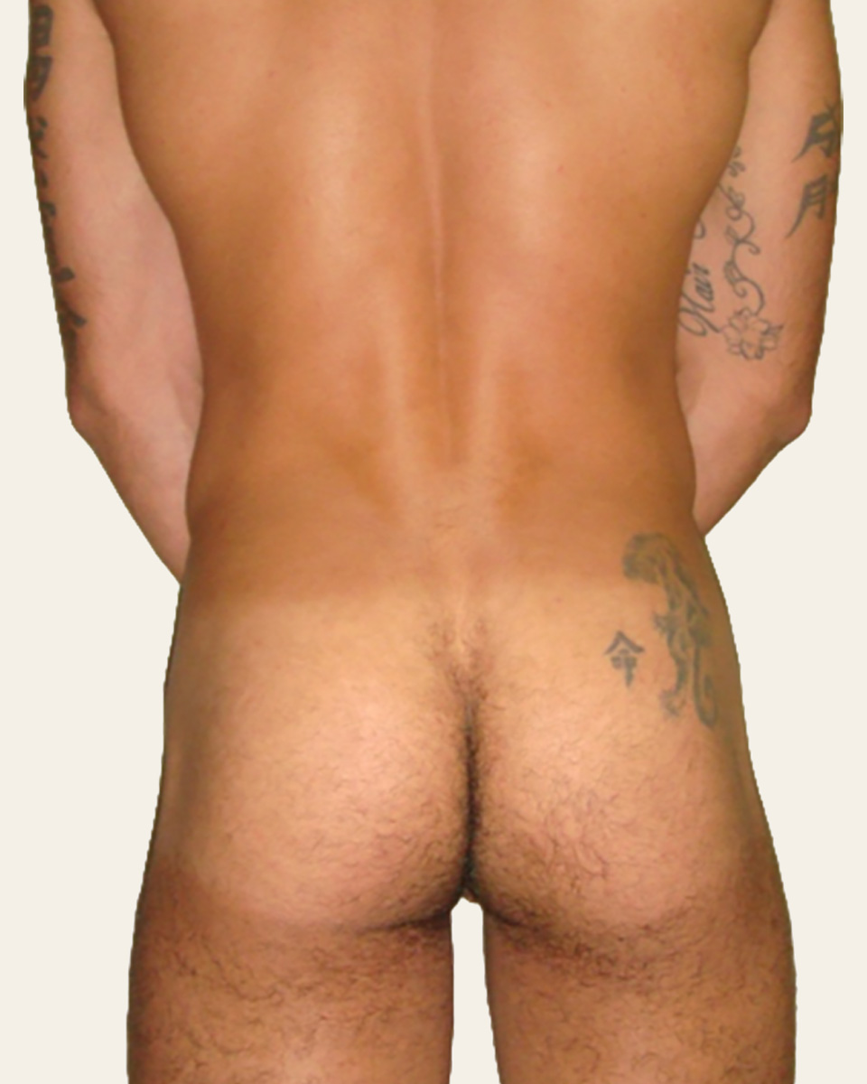
Clinical examples — individual anatomy and treatment objectives vary.
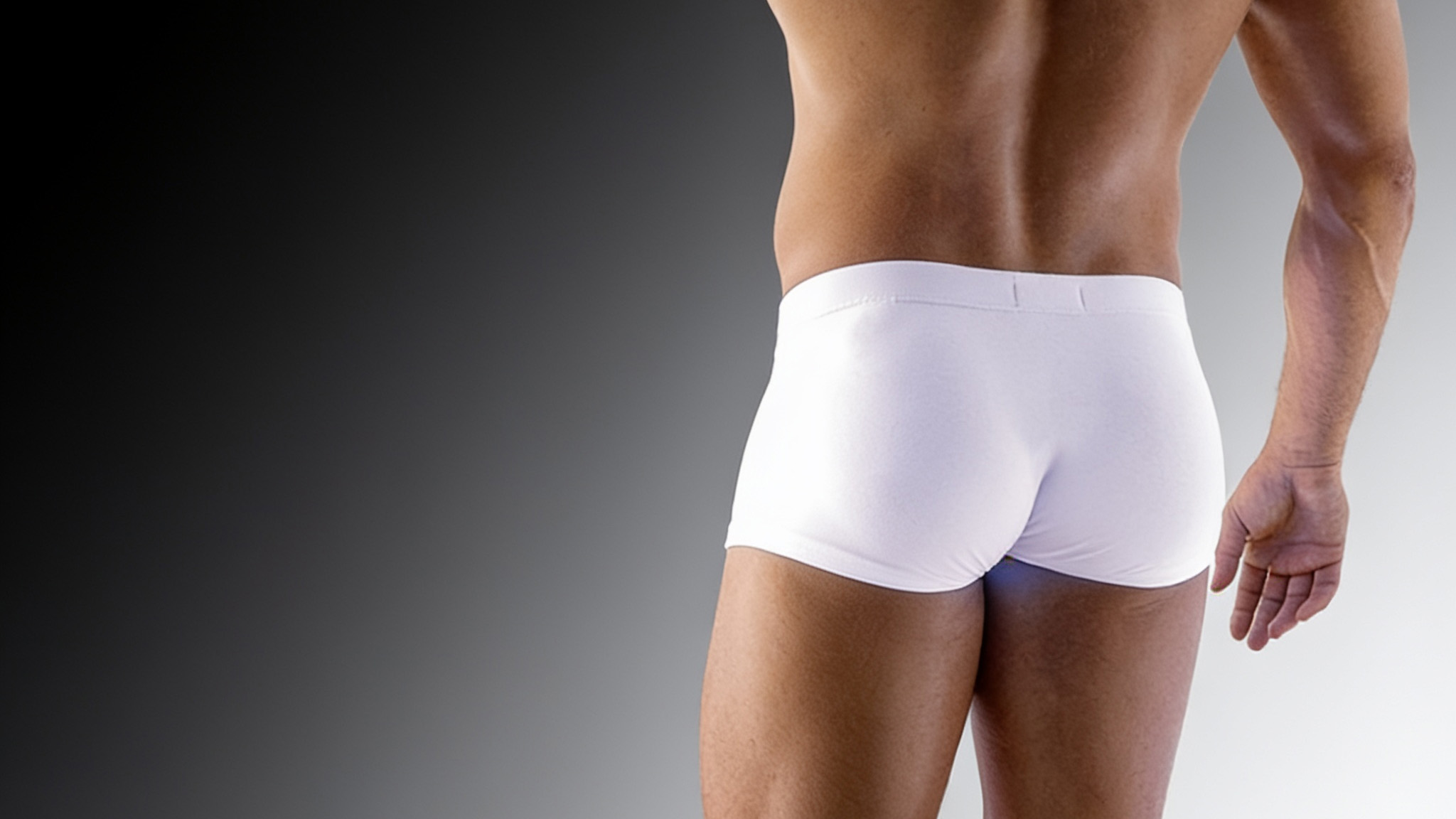
Athletic male gluteoplasty silhouette illustrating a compact, muscular gluteal contour

Male Gluteoplasty · Athletic Contour

Isovolumetric Reshaping for the Athletic Male Buttock

In athletic men, the objective is often not to add volume. Gluteal myomodulation reshapes an already developed muscular contour through a controlled lifting effect and a redistribution of the area of maximal projection.

Not augmentation. Redistribution.

Athletic gluteal hypertrophy may create substantial muscle mass while leaving the contour too vertically long, insufficiently compact, or poorly balanced in relation to the lumbar area and thighs. In these cases, adding volume would not necessarily improve the silhouette.

The treatment remains isovolumetric: no implant, filler or tissue addition is required. The intervention modifies the distribution of the existing muscular volume rather than increasing the total volume of the buttock.

Isovolumetric principle

Vertical length is reduced, width remains substantially unchanged, and the preserved volume is redistributed within a shorter contour. This can increase the local prominence of the area of maximal projection without creating an excessive result.
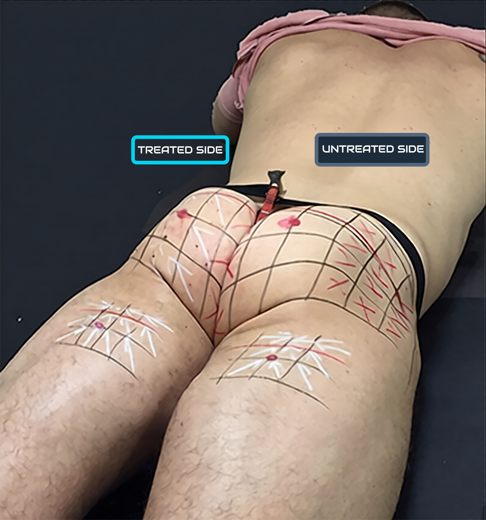
Treated and untreated side mapping for athletic male gluteoplasty Substantially preserved; the treatment primarily modifies vertical contour through a controlled downward translation and redistribution of the focused area of maximal projection.
L ↓ Vertical length Substantially preserved; the treatment primarily modifies vertical contour through a controlled downward translation and redistribution of the focused area of maximal projection.
W ≈ Transverse width Substantially preserved; the treatment primarily modifies vertical contour and the distribution of maximal projection.
V = Volume No added volume: an isovolumetric approach.
P ↑ Area of maximal projection More locally defined within the reshaped contour.
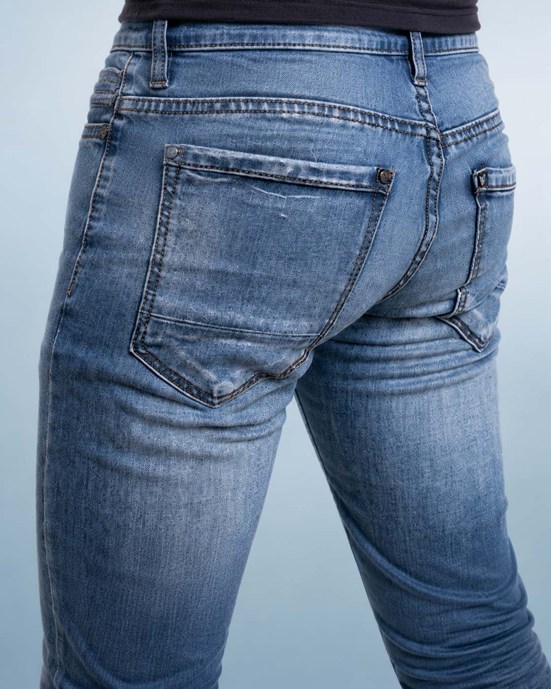
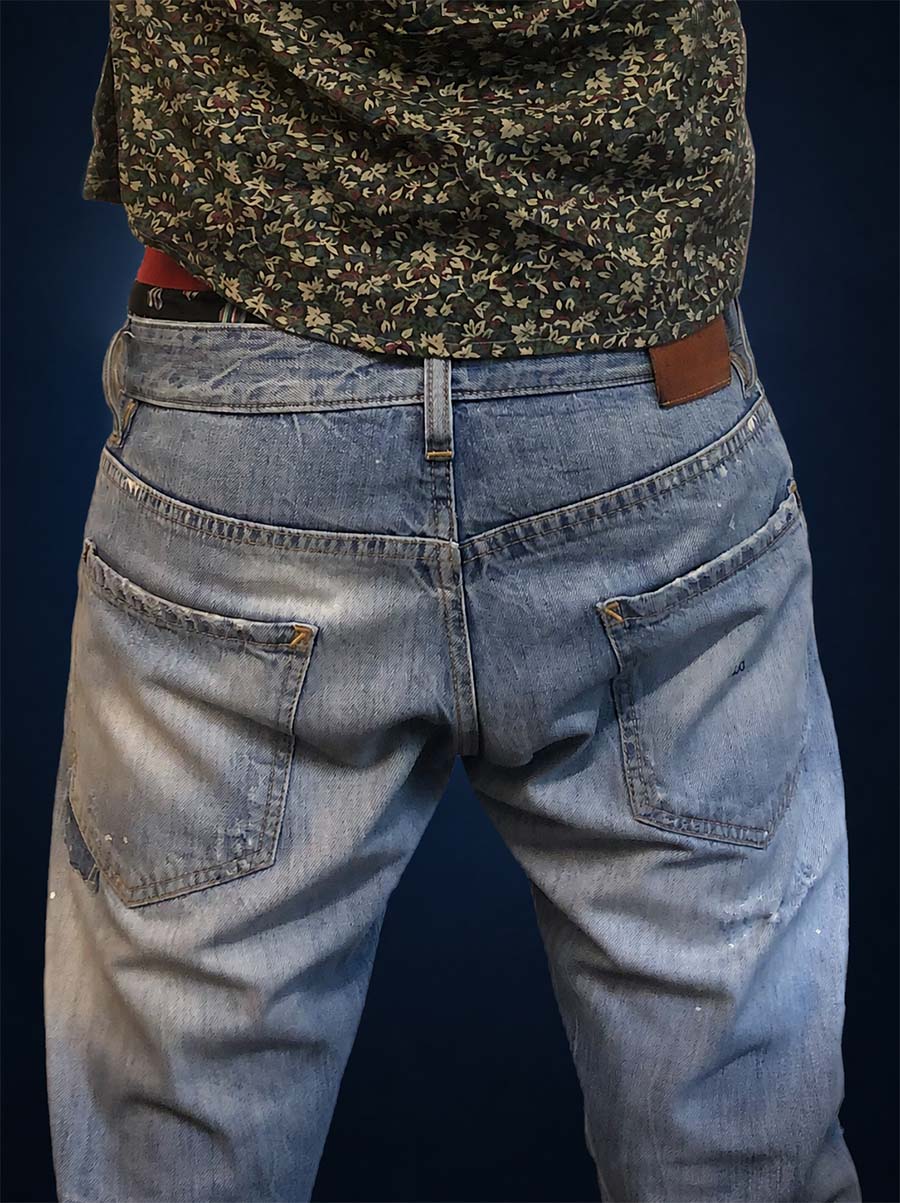
Athletic male low-rise jeans illustrating a compact lower gluteal contour

Aesthetic consequence

A lower area of maximal projection

The essential change is neither a wider buttock nor indiscriminate additional projection. It is the controlled relocation of the area of maximal projection within the lower gluteal region, while maintaining a natural masculine architecture.

Contemporary low-rise styling can visually emphasize this lower position of gluteal projection. The desired result remains compact, muscular and proportionate to the athlete’s body—not elongated.

Explore Male Gluteoplasty

Individual indications

Beyond a Single Aesthetic Template

Male gluteoplasty is not limited to one body type or one aesthetic goal. Planning is based on anatomy, muscular development, symmetry, tissue quality and the individual contour objective.

One method. Different clinical objectives.

The athletic contour has its own isovolumetric logic, but other patients may seek a change in shape, restoration of balance, correction of a secondary contour alteration or a more individualized expression of their body.

Myomodulation does not impose a standardized buttock shape. Each treatment plan is adapted to the relationship between the lumbar area, buttocks, thighs and the patient's overall silhouette.
Individualized assessment and planning for male gluteal contour reshaping
Before and after comparison of athletic male gluteal contour refinement
01

Athletic contour refinement

In men with developed gluteal musculature, the objective may be to create a more compact architecture through controlled lifting and a downward translation of the focused area of maximal projection.

Explore the athletic approach

Individual silhouette and gender-expression goals in gluteal contour planning
02

Shape and gender-expression goals

Some patients prefer a firmer, more angular and compact contour; others seek a softer, rounder or more projected silhouette. The intended result is discussed openly and planned within anatomical and proportionate limits.

Male gluteal asymmetry and contour irregularities
03

Asymmetry, atrophy and contour loss

Treatment may be considered when asymmetry, localized muscular atrophy, aging, weight variation or a previous procedure has altered the balance between the two sides or reduced definition of the gluteal-thigh transition.
Male gluteal implant contour before and after myomodulation
04

Secondary and reconstructive situations

Post-traumatic changes, sequelae of previous surgery or implants, and selected functional situations require a particularly careful medical assessment. Treatment is considered only when the anatomy, tissue condition and overall clinical context are appropriate.

The objective is never a template. It is a coherent contour, individualized to the patient's anatomy, movement and personal aesthetic identity.
Treatment options

Shape, Support and Function — Not Volume Alone

Male gluteoplasty should not be reduced to making the buttocks larger. Excessive or poorly positioned mass may increase visual heaviness, obscure muscular definition and create a lower, less coherent contour. Treatment should instead address architecture, support, projection, muscular balance and the relationship between the buttocks and the surrounding silhouette.

Male gluteal anatomy illustrating a structure-first approach to contour, muscular support and proportion
Male anatomical illustration — a structure-first approach. Effective planning considers muscular architecture, tissue support and overall proportion rather than simply adding more volume.
A structure-first strategy

More volume is not necessarily a better result

The buttocks are a dynamic anatomical structure, not an empty space that simply needs to be filled. Any added mass must be carried by the muscular framework, skin envelope and connective tissues. When it is excessive, poorly distributed or insufficiently supported, the result may appear heavy, descend visually and lose definition.

Planning therefore begins by identifying the true source of the contour problem: muscular position, excessive contraction, inadequate support, localized irregularity, implant-related distortion or an imbalance between the lower back, buttocks and thighs.

The objective is the smallest effective correction that restores a supported, proportionate and functional contour.

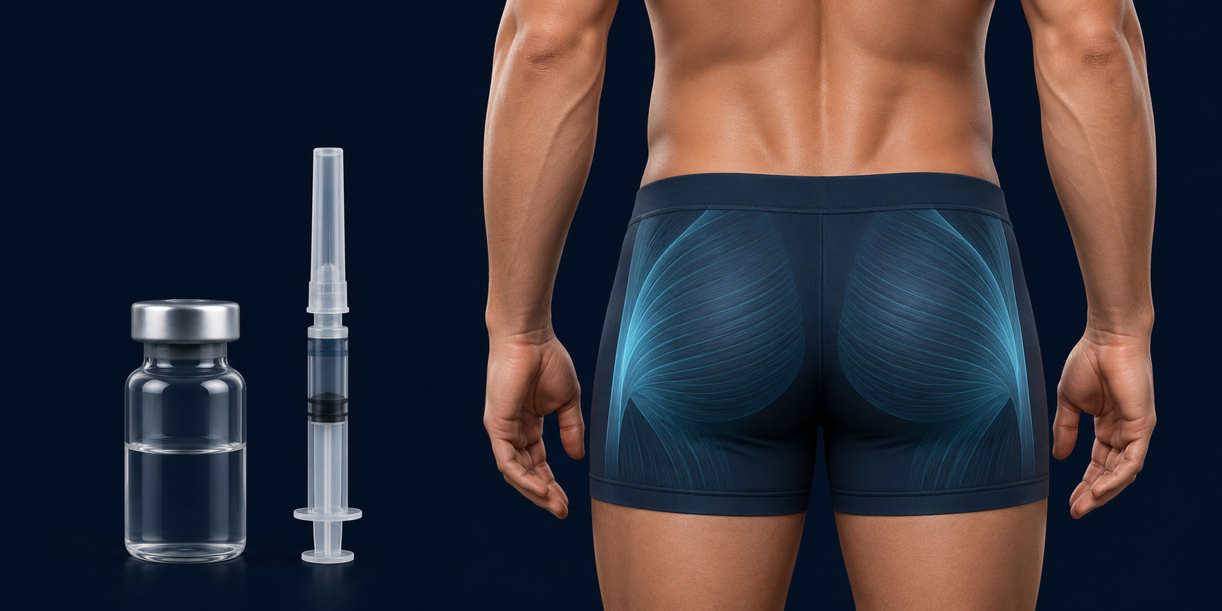
Male gluteal anatomy with a generic botulinum toxin vial and syringe, illustrating its limited non-volumizing role
02 Controversial
Neuromuscular inhibition

Botulinum toxin

Some practitioners use botulinum toxin to alter or round the gluteal contour through selective muscular relaxation. The visible change is obtained by reducing neuromuscular activity, not by strengthening or repositioning the muscular architecture.

Weakening an injected muscle may be accompanied by loss of muscle volume, particularly when treatment is repeated. Because the gluteal muscles contribute to movement, mass and dynamic support, routine cosmetic paralysis conflicts with a strategy intended to preserve strength and long-term structural quality.

Accelerated gluteal ageing has not been quantified in adequate long-term clinical studies; it remains a biological and clinical concern rather than a proven universal outcome.

Included for completeness — not presented here as a preferred contouring strategy.

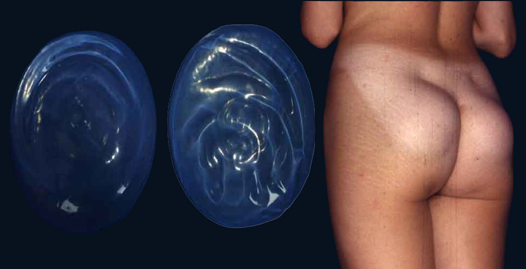
Gluteal implants beside a female clinical example illustrating implant-created projection
Female clinical example — implant-created projection Gluteal implants can create substantial projection, but projection alone does not guarantee harmonious integration with the pelvis and surrounding tissues. Implant dimensions, position and tissue coverage must be adapted to the individual anatomy.
03 Selected cases
Controlled structural projection

Implant-based procedures

Gluteal implants should not be understood as a routine way to make the buttocks larger. In carefully selected patients, they may provide defined projection that cannot be obtained through muscular treatment alone. Their dimensions, position and tissue coverage must be tightly controlled to limit excessive weight, visible distortion and an anatomically incoherent result.

Projection must remain proportionate to the tissues that support it.

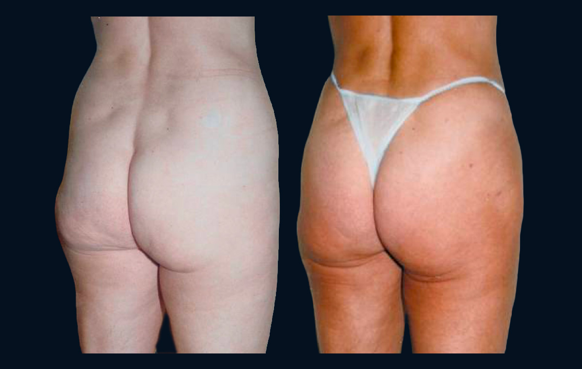
Female clinical comparison illustrating increased gluteal volume without equivalent improvement in structural support
Female clinical example — volume versus structural support Greater gluteal volume does not necessarily create a higher or better-supported contour. Added mass may increase projection while the lower pole remains low and the infragluteal folds remain pronounced. This non-standardized comparison is presented for qualitative anatomical illustration.
04 Conservative use
Localized refinement and proportions

Fat grafting, fillers and complementary contouring

Fat grafting, selected fillers and contouring of the surrounding areas may improve a localized depression or transition. They should be used as precise corrective tools rather than as a strategy of indiscriminate enlargement. Added mass can increase projection, but it does not create muscular support and may accentuate heaviness when the tissues are already descending.

Volume can alter size; it cannot replace structure.

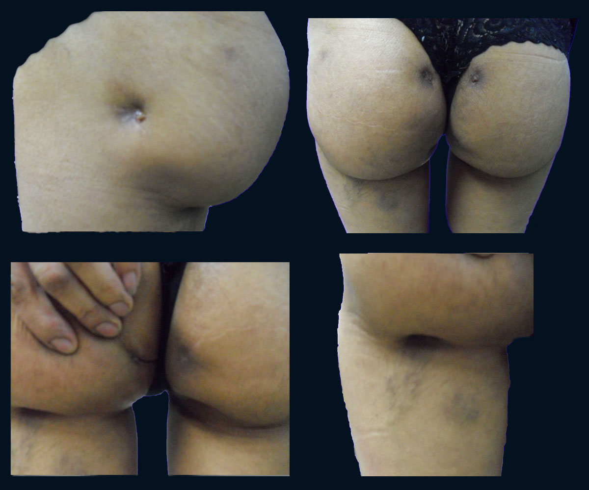
Four clinical views of localized gluteal contour depressions and tissue distortion in a female patient
Female clinical example — contour irregularities associated with gluteal threads Multiple views illustrate localized depressions and tissue distortion, including changes demonstrated during palpation. This reported clinical presentation does not represent every thread-based procedure.
05 Limited role
Temporary soft-tissue suspension

Gluteal threads

Suspension threads are proposed by some practitioners to create a modest lifting effect without surgery. They act on the superficial soft tissues; they do not reposition the gluteal muscle, rebuild its architecture or remove the weight that contributed to descent.

The buttocks are a heavy, highly mobile and load-bearing region. Expecting thin threads to provide durable structural support may therefore be mechanically unrealistic. Results may be limited or temporary, while irregularity, palpability, asymmetry, inflammation, infection, migration or extrusion are relevant concerns.

Techniques and materials vary, and robust long-term comparative evidence for durable gluteal lifting remains limited.

Shown because it is marketed as an option — not endorsed here as a structural treatment.

Gluteal Myomodulation

Dynamic Contour Assessment and Gluteal Myomodulation

Male gluteal contour is not purely static. Muscular contraction can reveal or accentuate contour variations that are less apparent at rest. Assessment should therefore consider the buttocks in both relaxed and contracted conditions, together with their relationship to the waist, hips and upper thighs.

Dynamic assessment

External Concavity at Contraction

A lateral gluteal concavity may become more visible when the gluteal muscles contract. In a masculine contour, this area contributes to the transition between the upper buttock and the lateral thigh.

Its presence alone does not establish an indication for treatment. Its location, depth, symmetry and behaviour during movement must be assessed individually before planning any intervention.

Clinical principle

The objective is not automatically to erase every depression or to add volume. It is to evaluate whether a selected contour relationship is compatible with the patient’s anatomy and desired masculine silhouette.
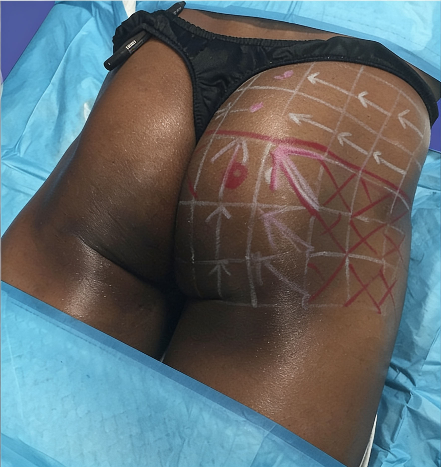
Male lateral gluteal contour with surface markings used for clinical assessment and treatment planning
Male clinical example — mapping of the lateral gluteal contour. Surface markings document the anatomical areas considered during clinical assessment and treatment planning. They do not, by themselves, identify the procedure performed or demonstrate a clinical result.

Treatment principle

Not Augmentation. Selective Dynamic Reshaping.

Gluteal myomodulation is intended to influence selected muscular and contour relationships rather than simply increase gluteal volume. Its planning should preserve a coherent transition between the lower back, buttock and thigh, while respecting the patient’s existing muscular development.

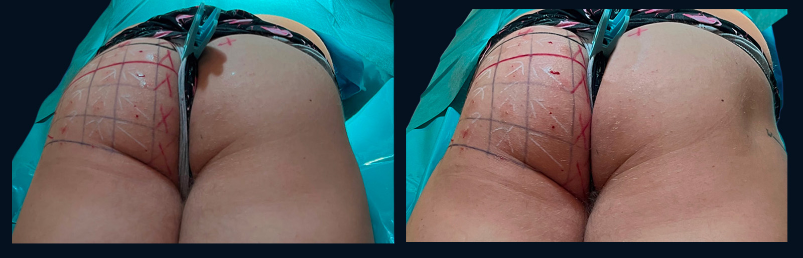
Early unilateral clinical observation

Relaxation and Contraction, 30 Minutes After Treatment

The following paired views document an immediate unilateral observation under relaxed and contracted conditions.

Early unilateral clinical observation

Relaxation and Contraction, 30 Minutes After Treatment

The following paired views document the same unilateral clinical observation under relaxed and contracted conditions. Each condition is shown from two complementary viewing angles.
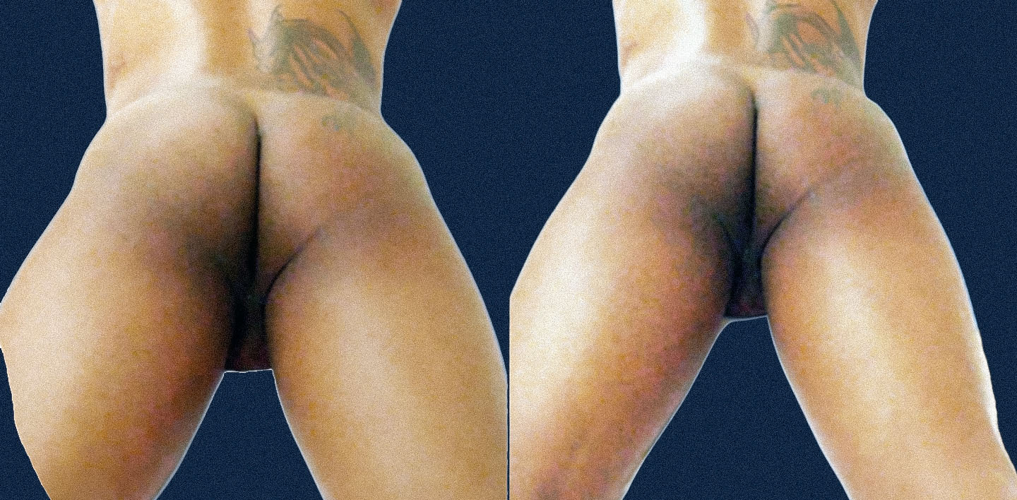
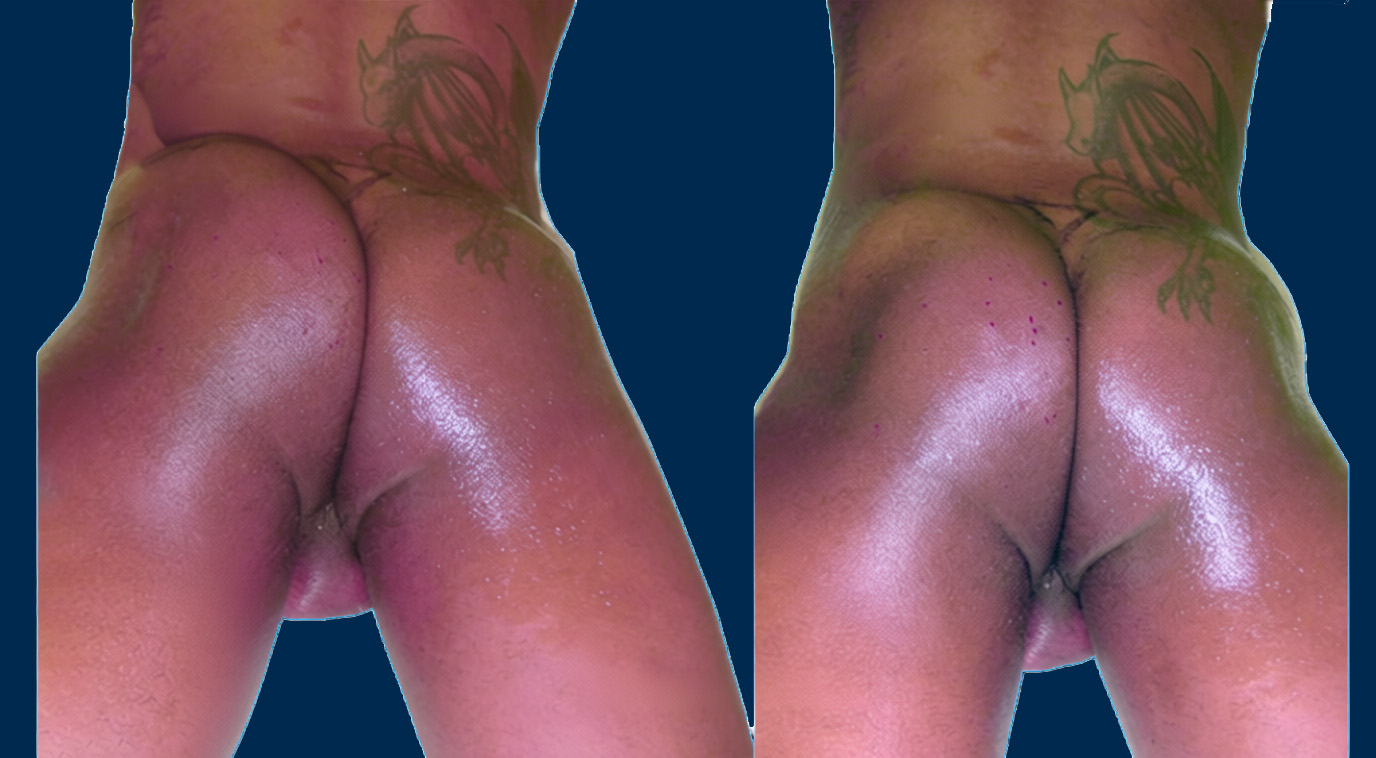
Relaxation
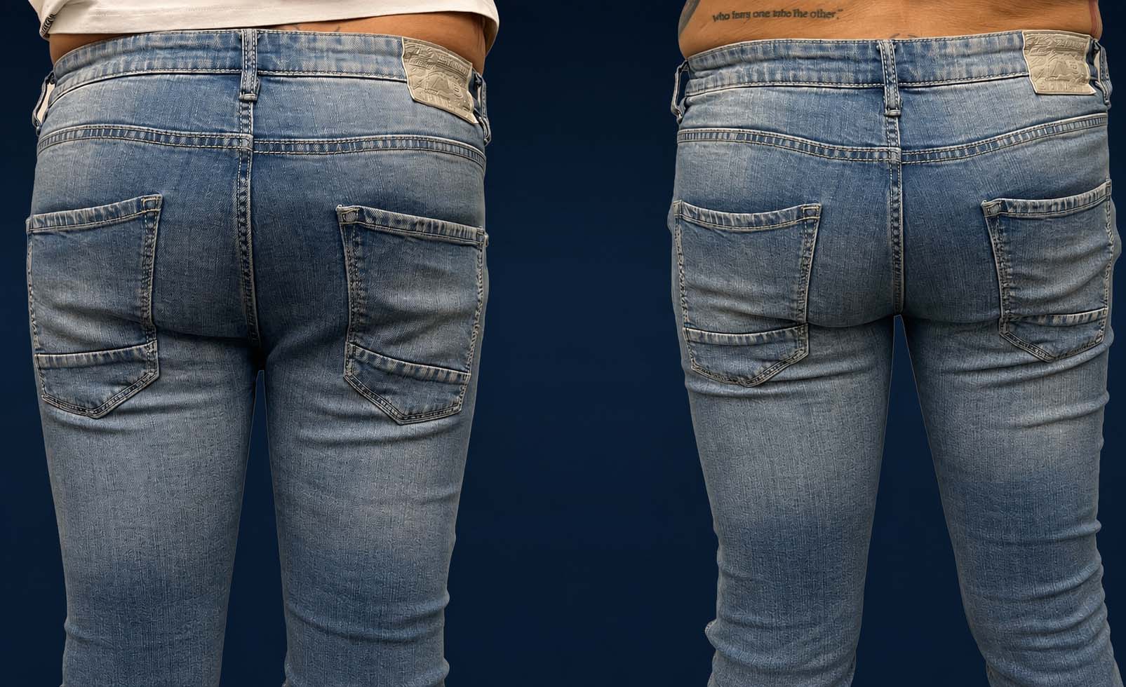
Male gluteal contour at relaxation 30 minutes after unilateral myomodulation, shown from two complementary viewing angles, with the patient's left side treated and right side untreated
Relaxation — immediate unilateral comparison from two viewing angles. Both photographs show the same patient under relaxed conditions. Only the patient’s left gluteal side was treated; the right side remained untreated for immediate comparison.
Contraction
Male gluteal contour during contraction 30 minutes after unilateral myomodulation, shown from two complementary viewing angles, with the patient's left side treated and right side untreated
Contraction — immediate unilateral comparison from two viewing angles. Both photographs show the same patient during muscular contraction. Only the patient’s left gluteal side was treated; the right side remained untreated for immediate comparison.

These individual early observations do not establish the durability or typical magnitude of a result. Images courtesy of Dr Alain Tenenbaum.

Clinical planning

Patient-Specific Assessment & Treatment Mapping

There is no universal marking pattern. Clinical assessment determines the treatment map for each patient according to the existing anatomy, muscular behaviour, previous procedures and the contour the patient wishes to preserve or modify.

The clinical principle

The Intended Contour Must Be Defined Before Marking

The position of maximum projection, the intergluteal region and the external gluteal concavity must be assessed together. Modifying one component may change the visual relationship between projection, width and the surrounding anatomical transitions.

The objective is not to reproduce a standardised shape. The treatment map is adapted to the patient’s baseline morphology, asymmetries, dynamic muscular response and stated contour preferences.

The marking is designed for the individual patient—not copied from a fixed template.
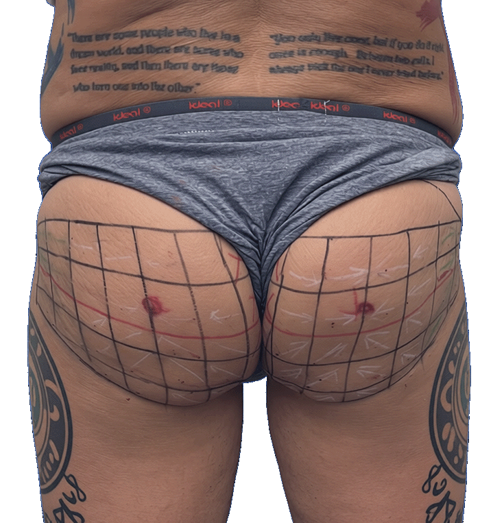
Patient-specific gluteal treatment marking showing maximum projection points and individualized anatomical mapping
Patient-specific treatment mapping in a male patient with existing gluteal implants, showing maximum projection points and side-specific anatomical markings.

Contour preference

Define the Intended Contour

Assessment begins by identifying what the patient wishes to preserve, emphasise or soften. General labels alone are insufficient; the desired anatomical changes should be described precisely.

  • Preserve, reinforce or visually reposition the point of maximum projection
  • Preserve the intergluteal region or seek a visually broader appearance
  • Maintain or accentuate the external concavity
  • Soften or fill the external concavity when a rounder transition is preferred
  • Preserve a typically masculine contour or pursue selected feminising contour characteristics

Clinical examination

Assess the Individual Anatomy

Static appearance alone does not provide sufficient information. Examination at rest and during voluntary contraction helps identify asymmetries and the apparent contribution of muscular activity to the visible contour.

  • Posterior, oblique and lateral contour relationships
  • Position of maximum gluteal projection
  • Static and dynamic right-to-left asymmetries
  • External concavity and lateral transitions
  • Skin quality and distribution of the soft tissues
  • Muscular behaviour at rest and during contraction

Individual mapping

Adapt the Marking and Treatment Plan

The findings are translated into a side-specific treatment map. Existing procedures or anatomical constraints may substantially modify the marking, the proposed approach or the feasibility of treatment.

  • Adapt the marking independently to each side
  • Account for existing gluteal implants
  • Identify previous injections or transferred material
  • Consider previous surgery, scars or tissue alteration
  • Determine whether another strategy is more appropriate
  • Avoid treatment when safe planning cannot be established

Interdependent planning points

Three Contour Elements Must Be Considered Together

The treatment map should reflect the relationship between projection, central definition and the external contour rather than treating each area as an isolated feature.

Maximum Projection Point

Its baseline position is documented before deciding whether the visual emphasis should be preserved, reinforced or rebalanced.

Intergluteal Region

The patient’s preference for preserving or visually broadening this region is considered in relation to the surrounding contour.

External Gluteal Concavity

The plan may seek to preserve or emphasise the concavity, or to soften it when the patient prefers a fuller lateral transition.

Assessment before treatment

A Visible Contour Concern Does Not Define Its Cause

Clinical assessment comes before treatment selection. It determines whether the patient’s objective is compatible with the anatomy, whether a patient-specific marking can be established and whether the proposed approach is appropriate.

General and gluteal-area-specific contraindications are addressed separately and remain part of the medical safety assessment.

Review Contraindications

Treatment pathway

Treatment Journey, Results & Follow-Up

Gluteal reshaping is a progressive clinical process. Each session is followed by standardised assessment so that immediate changes, subsequent evolution and the need for further treatment can be evaluated separately.

15–30 minutes

Approximate session duration

None required

No anaesthesia is routinely used

30 minutes

Immediate contour change may already be visible

3 days

First reasonable clinical assessment of the result

Before treatment

Document the Baseline Contour

Clinical examination, patient-specific marking and standardised photography establish the reference against which subsequent changes will be assessed.

  • Posterior, oblique and lateral photographs with consistent positioning, distance and lighting
  • Additional views at rest and during voluntary gluteal contraction when dynamic assessment is relevant
  • Documentation of the point of maximum projection, external concavity and intergluteal region
  • Assessment in the patient’s preferred jeans with back pockets when the clothed contour forms part of the aesthetic objective

Treatment session

A Short Outpatient Procedure

The treatment is performed on an outpatient basis according to the patient-specific map established during clinical assessment. A session generally lasts approximately 15 to 30 minutes.

Outpatient treatment No anaesthesia Patient-specific marking

Approximately 30 minutes

Document the Immediate Change

A visible modification of the contour may be observed immediately and documented approximately 30 minutes after treatment. These photographs provide an early comparison, but they should not be confused with the subsequent clinical assessment.

The 30-minute photographs document an immediate contour change. They do not represent the final long-term result.

After three days

First Clinical Evaluation of the Result

The result can be evaluated more reasonably after approximately three days. Comparison with the baseline photographs helps assess projection, width, anatomical transitions and right-to-left balance.

Results remain patient-specific and depend on the initial anatomy, muscular behaviour, treatment map and individual clinical response.

Longitudinal follow-up

Compare, Review and Adapt

Follow-up is used to compare standardised photographs, examine the contour at rest and during contraction, and decide whether the treatment schedule should be maintained or adapted.

Clinical reviews may be scheduled at approximately 1, 3, 6 and 12 months. A follow-up visit does not necessarily correspond to an additional treatment session.

Static and dynamic assessment

Results Are Compared at Rest and During Contraction

Standardised posterior, oblique and lateral photographs help document changes in projection, lifting and contour from several complementary viewing angles.

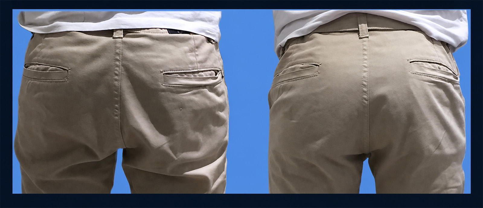
When muscular behaviour contributes to the visible contour, the same views are reproduced at rest and during voluntary contraction. Assessment with the patient wearing his preferred jeans may also show how the treatment changes the clothed silhouette.
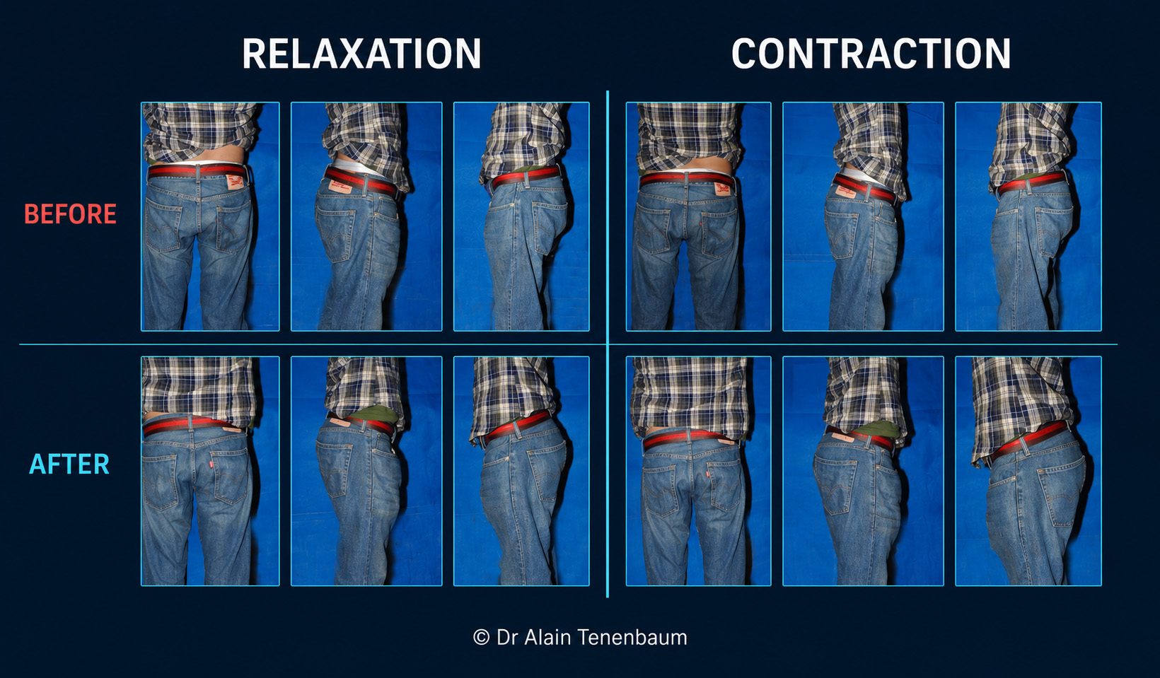
Male gluteoplasty results compared before and after treatment in posterior, oblique and lateral views, at rest and during muscular contraction
Standardised before-and-after views document the male gluteal contour at rest and during voluntary contraction from posterior, oblique and lateral angles.

Progressive treatment schedule

Five Sessions During the First Two Years

The initial treatment protocol is distributed over two years rather than being concentrated into a single period. The exact timing may be adapted according to the patient’s clinical response and objectives.

Year 01 3

Treatment Sessions

Three patient-specific sessions are generally planned during the first year.

Year 02 2

Treatment Sessions

Two additional sessions generally complete the initial two-year protocol.

Maintenance 2

Sessions Per Year

After the initial phase, maintenance is generally performed twice per year, at approximately six-month intervals.

Follow-up appointments and treatment sessions are separate clinical events. A review does not automatically result in another treatment.
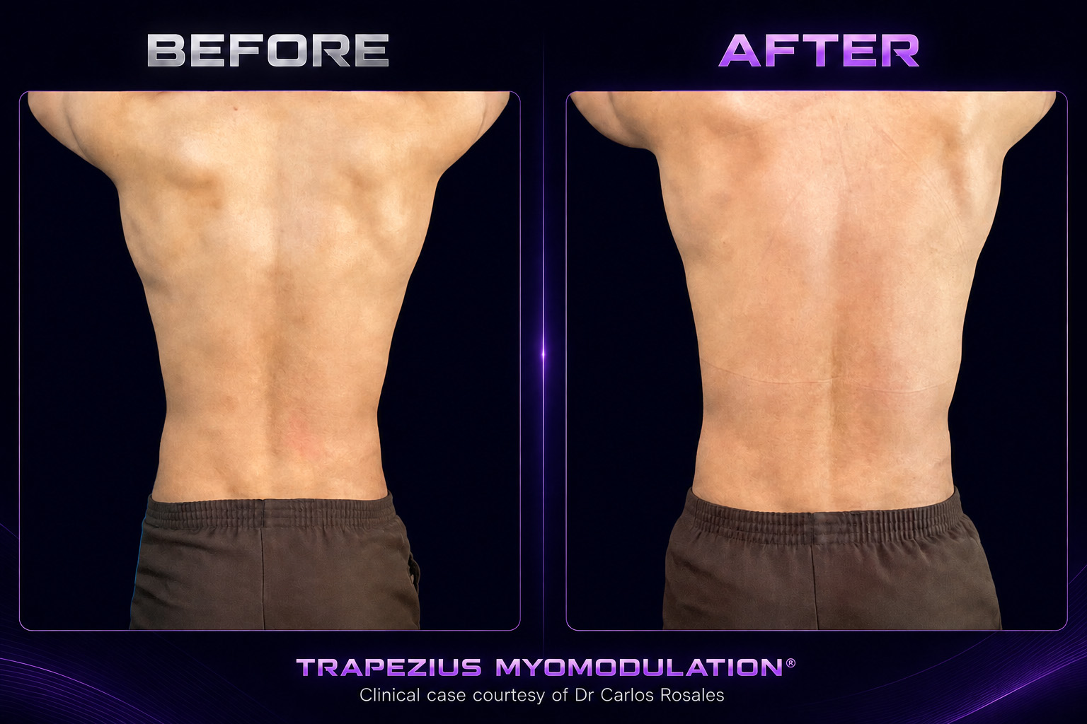
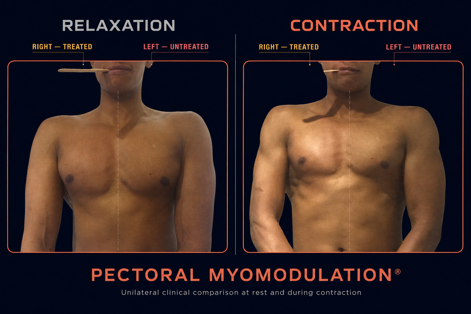
Patient-specific results

Results Must Be Assessed Over Time

Immediate visibility does not replace longitudinal assessment. Reproducible photographs and clinical examination allow the practitioner to distinguish the early contour change from its subsequent evolution and to adapt the protocol when necessary.

View Clinical Results

Clinical safety

Safety, Limitations & Previous Procedures

Safety depends not only on the visible contour, but also on what may already exist beneath it. Implants, injected materials, fat transfer, scars and previous surgery can change the examination, imaging requirements, marking and feasibility of treatment.

The clinical principle

A Technically Possible Treatment Is Not Always Appropriate

A reliable treatment map can only be established after complete disclosure of previous procedures and careful clinical assessment. Depending on the findings, treatment may proceed with an adapted plan, require imaging, be postponed or be avoided.

What may already be present

Previous Procedures Require Individual Assessment

The type of procedure, material used, date, anatomical plane and current tissue condition should be documented whenever possible.

Gluteal Implants

A correctly positioned implant does not automatically exclude treatment. Feasibility is assessed case by case after clinical examination, MRI and ultrasonography, with a modified treatment map when appropriate.

A superficially positioned implant is an absolute contraindication until the implant has been removed.

Previous Fat Transfer

Previous gluteal fat transfer may remain compatible with treatment because transferred fat is vascularised. Clinical examination is nevertheless required before marking and treatment planning.

Resorbable Fillers

The injected product, anatomical location and date should be identified whenever possible. Incomplete information may require additional assessment before treatment is considered.

Surgery and Scars

Previous surgery and scars may alter tissue planes and muscular behaviour. Treatment is generally postponed for six to twelve months, then reconsidered according to healing and anatomy.

Lumbar Dermolipectomy

Previous lumbar dermolipectomy, including after major weight loss, is not necessarily a permanent contraindication. It requires individual reassessment of scars, tissues and anatomical changes.

Previous Biopolymers

Previous biopolymer injections require enhanced assessment and specific informed consent. Pre-existing biopolymers may migrate or cause delayed inflammatory complications independently of the proposed treatment.

Any subsequent change must be clinically evaluated rather than automatically attributed to either the previous material or the myomodulation procedure.

Different reasons not to proceed

Safety Restrictions and Treatment Limitations Are Not the Same

Some findings create a safety contraindication, some require a delay, and others mean that a visible result is unlikely even when treatment could technically be performed.

Do not treat

Absolute Contraindications

  • Superficially positioned gluteal implant
  • Dangerous copies or unverifiable products
  • Copied or unauthorised hydroxybenzene products
  • Peptones alone or in combination

Postpone and reassess

Temporary Restrictions

  • Anabolic steroid use within the previous three months
  • Recent surgery or incompletely healed scars
  • Missing information about a previous injected product
  • Any active local condition requiring medical assessment

No meaningful visible benefit

Effectiveness Limitation

When a substantial layer of gluteal fat covers the muscle, the muscular contour remains hidden. Treatment is therefore not appropriate because a visible result should not be expected.

After treatment

Expected Reactions Must Be Distinguished From Complications

Small red marks at injection points and ecchymoses may occur. An ecchymosis is a superficial bruise and should not be confused with a haematoma. Allergy is reported very rarely.

Pain, tenderness, haematoma and swelling are not routinely expected. Any unexpected, persistent or worsening reaction requires prompt assessment by the treating practitioner.

May occur Red injection-point marks
May occur Ecchymoses
Very rare Allergic reaction
Seek assessment Unexpected or worsening symptoms

Complete medical information

Review the Dedicated Safety Pages

This section highlights considerations specific to treatment planning. It does not replace a complete medical consultation or the detailed information on contraindications, side effects and complications.

Clinical FAQ

Questions before treatment

Male Gluteoplasty Frequently Asked Questions

Concise answers about treatment planning, results, recovery, previous procedures and the progressive maintenance protocol.

01 What is male gluteal myomodulation designed to change?

Treatment is designed to modify the visible relationship between gluteal projection, width, lifting and anatomical transitions by acting according to a patient-specific muscular treatment map. It is not based on a universal male shape or a standard marking pattern.

02 Is the marking the same for every patient?

No. The position of maximum projection, intergluteal region, external concavity, asymmetries and muscular behaviour are assessed together. The marking is adapted independently to each side and to the contour the patient wishes to preserve or modify.

03 How long does one treatment session take?

A session generally lasts approximately 15 to 30 minutes. It is an outpatient treatment and no anaesthesia is routinely required.

04 When can a change be seen and assessed?

A visible contour change may already be documented approximately 30 minutes after treatment. The first reasonable clinical assessment is performed after about three days. Immediate images should not be presented as the final long-term result.

05 Can normal activities and exercise be resumed immediately?

Normal daily activities and exercise may generally be resumed immediately. No routine restriction concerning sitting, massage or voluntary muscular contraction is required.

06 How many sessions are included in the protocol?

The initial protocol generally includes five sessions over the first two years: three during the first year and two during the second. After this initial phase, two maintenance sessions per year are generally planned, usually at approximately six-month intervals.

07 Why are results photographed at rest and during contraction?

The visible contour may change with muscular activity. Standardised posterior, oblique and lateral photographs at rest and during contraction help document dynamic behaviour, asymmetries and the evolution of projection and lifting.

08 Can a patient with gluteal implants be treated?

A correctly positioned implant may be considered on a case-by-case basis after clinical examination, MRI and ultrasonography. A superficially positioned implant is an absolute contraindication until it has been removed.

09 What if fillers, fat transfer or biopolymers were previously injected?

Previous fat transfer may remain compatible with treatment after examination. For resorbable fillers, the product and injection date should be documented whenever possible. Biopolymers require enhanced assessment and specific informed consent because they may migrate or cause delayed complications independently of the proposed treatment.

10 Can treatment be performed after surgery?

Previous surgery and scars require reassessment because tissue planes and muscular behaviour may have changed. Treatment is generally postponed for six to twelve months and reconsidered after healing. Previous lumbar dermolipectomy is not necessarily a permanent contraindication.

11 When is treatment unlikely to produce a visible result?

When a substantial layer of gluteal fat covers the muscle, the muscular contour remains hidden. In that situation, a meaningful visible result should not be expected even if treatment could technically be performed.

12 Can copied products, hydroxybenzene products or peptones be used?

No. Counterfeit or copied products, copied or unauthorised formulations described as containing hydroxybenzene (hidroxibenceno), peptones and any unverifiable product are absolute contraindications. Peptones must not be used alone or in combination. These products present their own safety risks.

Individual assessment

Still Have a Patient-Specific Question?

A consultation is required to relate the treatment objective to the patient’s anatomy, previous procedures and medical context.

Contact a Specialist