Clinical Results Atlas · 01 · Myomodulation®

Clinical Results Atlas

From clinical indication to three-dimensional outcome.

Gluteal Myomodulation® · Isovolumetric reshaping

A clinical reading of projection, lifting, lateral contour, symmetry and muscular dynamics. Existing cases are presented with their treatment objective, photographic interval and interpretation limits.

30 MIN

Early reconstructive observation

One post-traumatic atrophy case, photographed 30 minutes after one session.

1 H

Immediate contour observations

The other immediate case intervals are confirmed by the clinical author. Follow-up series retain their image-labelled dates.

Clinical Results Atlas · 1.3 · Myomodulation®

How to Read This Atlas

Begin with the indication. Read the anatomy, the view and the time point before interpreting the visible change.

01

Projection & Pmax

Profile views describe posterior projection; posterior views describe width, symmetry and contour. Follow the area of maximal projection in relation to fixed anatomical references.

02

Lifting & lower contour

Compare gluteal length, the banana fold and the infragluteal transition. A change in clothing or camera angle can alter their apparent position.

03

Rest & contraction

Compare rest with rest, and contraction with contraction. Different muscular states answer different clinical questions.

04

Patient’s right and left

Side labels refer to the patient’s anatomy. They must not be inferred solely from the left or right side of the photograph.

05

Time after treatment

30 minutes and 1 hour are immediate observations. The selected Swiss follow-up images separately show 7 days and 1 month.

06

Clinical context

A photograph illustrates one person at one time. It cannot alone measure volume, muscular strength, patient satisfaction or long-term stability.

Clinical Results Atlas · 02 · Myomodulation®

What Constitutes a Clinical Result?

A useful result connects a visible contour change to a defined clinical question.

INDICATION

Why was treatment considered?

Identify the contour concern and baseline anatomy before describing the result.

OBJECTIVE

What was intended to change?

Describe projection, lifting, lateral transition, symmetry or dynamic behaviour, rather than using size alone.

COMPARISON

What do the photographs show?

Specify the treated side, viewing angle, muscular state, session count and photographic interval.

This atlas is an educational collection of selected cases. It is not a controlled clinical trial, an estimate of typical results or a guarantee of an individual outcome.

Clinical Results Atlas · 03 · Myomodulation®

The Isovolumetric Results Framework

Read the contour through length L, transverse width W and height / posterior projection H.

L × W × H · V = constantA simplified spatial framework for reshaping existing contour.
L

Length & lifting

Describe the vertical extent and position of the lower contour.

W

Width & lateral contour

Describe the transverse contour and continuity with the hips and thighs.

H

Height / projection

Describe posterior expression and the position of maximal projection.

Myomodulation® uses the specific Endopeel® formulation. Endopeel® is a formulation used for Myomodulation®, not a product category or a volume-creating treatment. The isovolumetric objective is reshaping the existing contour.

L × W × H is a conceptual model, not a volumetric measurement of the irregular buttock shape. Photographs alone cannot verify exact volume conservation.

Clinical Results Atlas · 04 · Myomodulation®

Female Projection & Lifting

Complementary views help distinguish posterior projection from the position of the lower gluteal contour.

F01Result 1 h after treatment

Female projection and lifting · Latino case

Female gluteal projection and lifting, profile comparison
Profile view · treated side: Left · original clinical montage.
Female gluteal projection and lifting, posterior comparison
Posterior view · 1 session · original clinical montage.
Indication
Projection and lower-contour refinement
Objective
Isovolumetric projection and lifting
Treated side
Left (profile view)
Sessions
1 (posterior comparison)
Photographic interval
Result 1 h after treatment
Assessment
Posterior / profile as shown
Interpretation limits
Individual early observation. Posture, lighting and photographic angle may influence the apparent contour. This comparison does not quantify volume or establish durability.
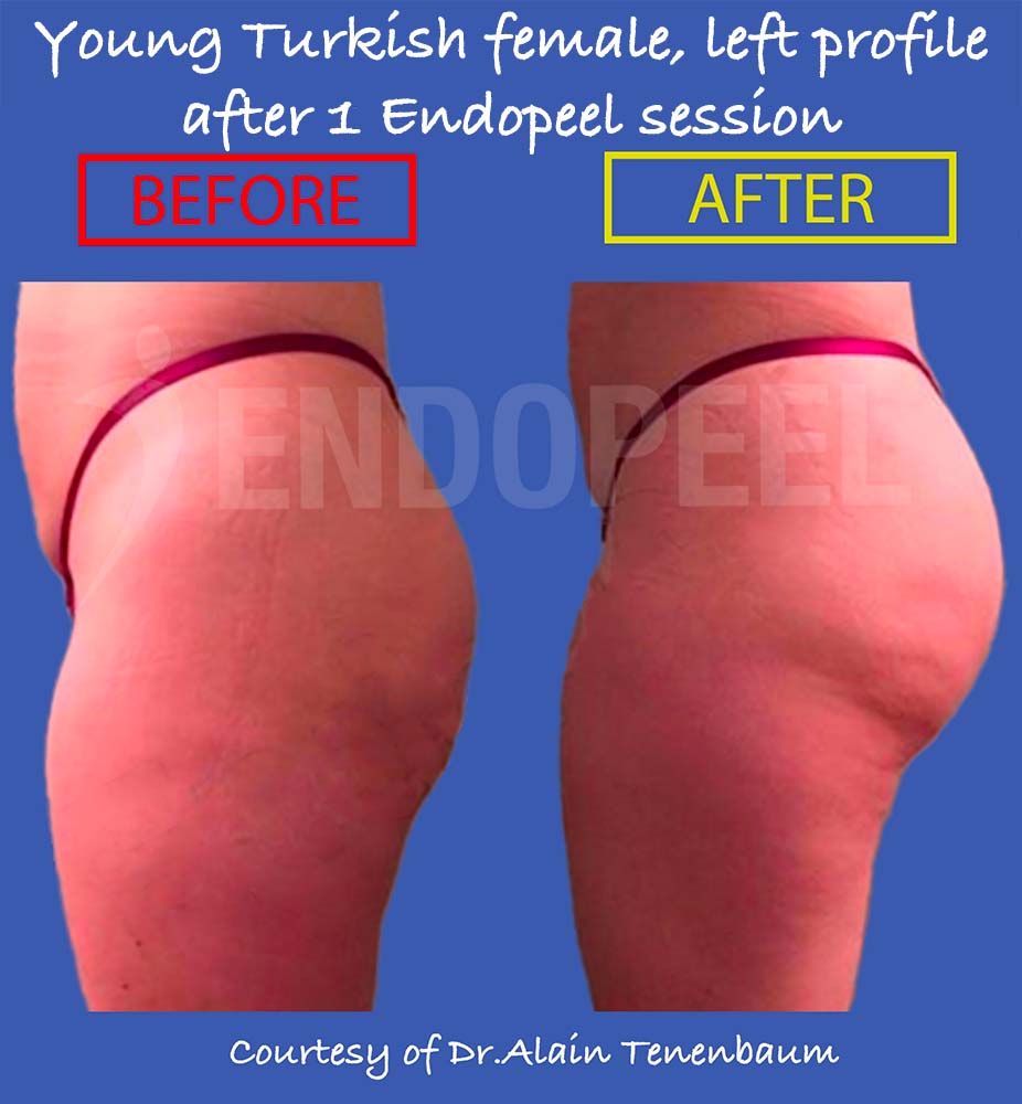
F02Result 1 h after treatment

Female profile refinement · Turkish case

Turkish female gluteal profile before and after treatment
Left-profile comparison · treated side: Left · 1 session.
Indication
Profile projection
Objective
Controlled posterior contour refinement
Treated side
Left
Sessions
1 session
Photographic interval
Result 1 h after treatment
Assessment
Left profile
Interpretation limits
Individual early observation. Posture, lighting and photographic angle may influence the apparent contour. This comparison does not quantify volume or establish durability.
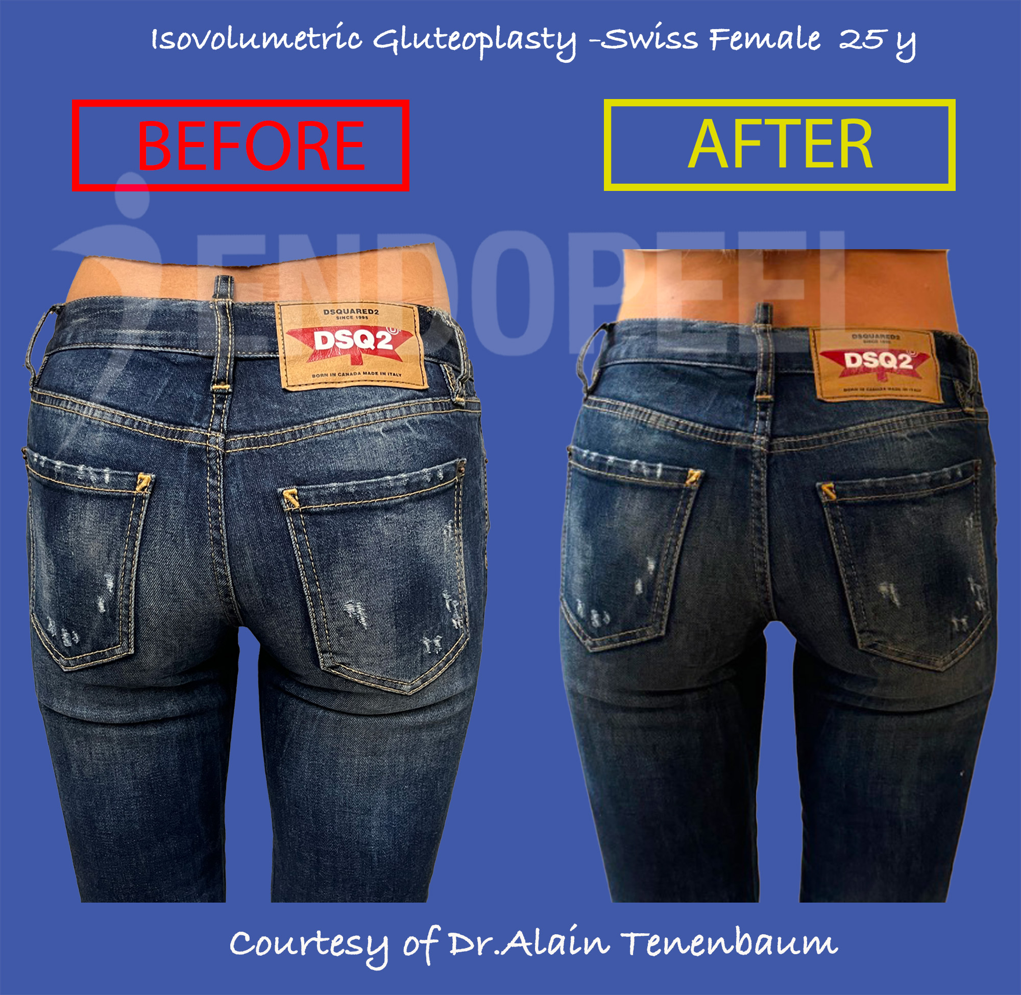
F03Result 1 month after treatment

Female gluteal projection · Swiss case

Swiss female posterior gluteal comparison
Posterior comparison.
Swiss female left-profile gluteal comparison
Left-profile comparison.
Indication
Projection and contour balance
Objective
Assess the reshaped contour in complementary views
Treated side
Both
Sessions
2
Photographic interval
Result 1 month after treatment
Assessment
Posterior and left profile
Interpretation limits
Clothing, pose and viewing angle may influence the apparent contour. This one-month comparison does not quantify volume or establish long-term durability.

Clinical Results Atlas · 05 · Myomodulation®

Latinisation & External Contour

Here, “Latinisation” describes an individual contour objective: lateral fullness, posterior expression and the relationship with the intergluteal region.
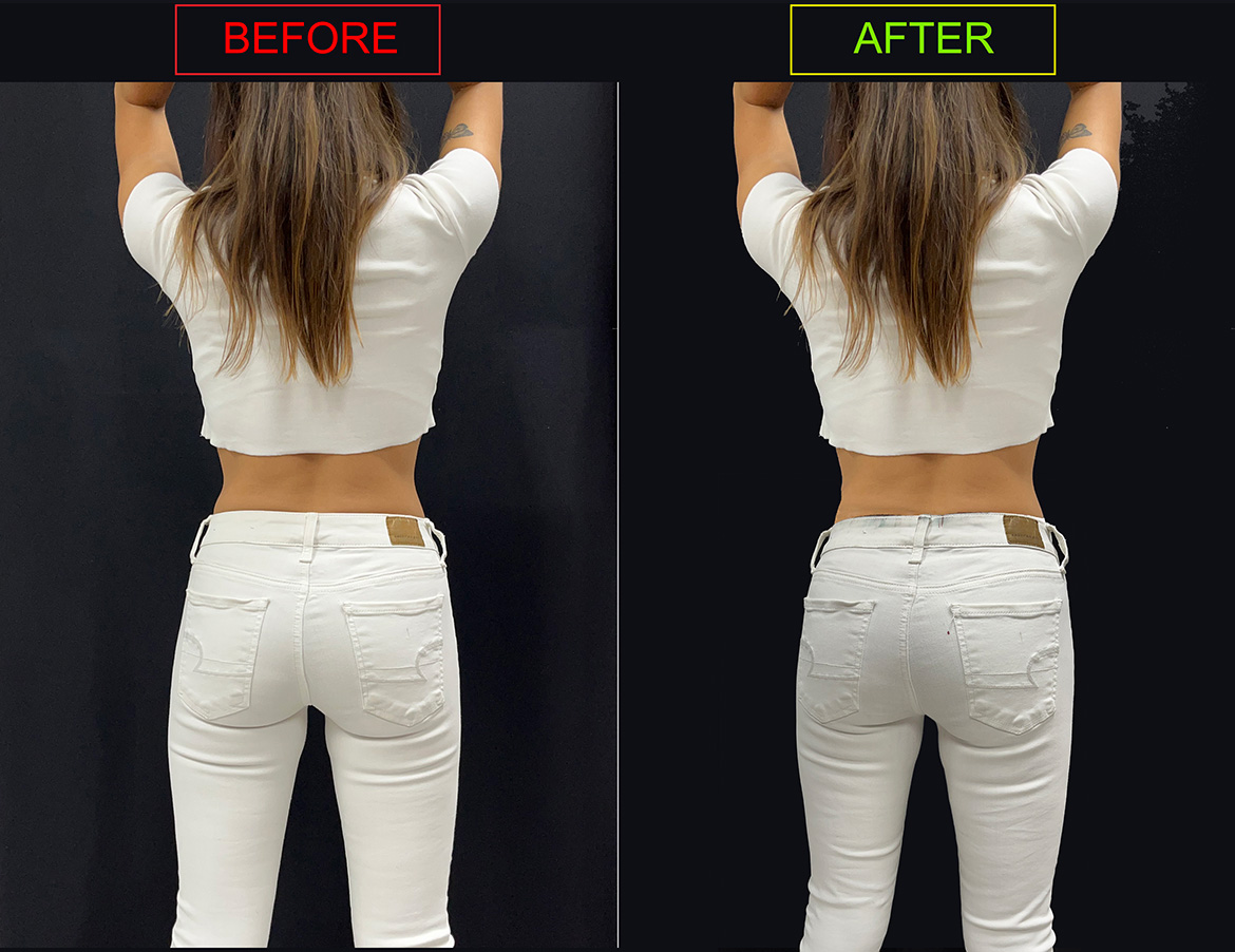
F04Result 1 h after treatment

Mexican female contour · individualized Latinisation

Mexican female gluteal contour comparison
Existing clinical comparison reused from Fundamental Basics.
Mexican female gluteal contour in white jeans
Clothed contour illustration from the original gallery.
Indication
Lateral contour refinement and widening of the intergluteal cleft
Objective
Discrete external Latinisation, projection, lower-contour lifting, widening of the intergluteal cleft and widening of the infragluteal diamond
Treated side
Both
Sessions
1
Photographic interval
Result 1 h after treatment
Assessment
Clinical comparison and clothed posterior contour
Interpretation limits
The case illustrates widening of the intergluteal cleft and infragluteal diamond, projection, banana-fold / infragluteal lifting and discrete external Latinisation. Clothing tension and pocket geometry can influence the visible change. A cultural label does not prescribe one ideal body shape.

Clinical Results Atlas · 06 · Myomodulation®

Unilateral Treatment as Internal Control

A treated side and an untreated side in the same patient offer a useful internal comparison at the same time point.
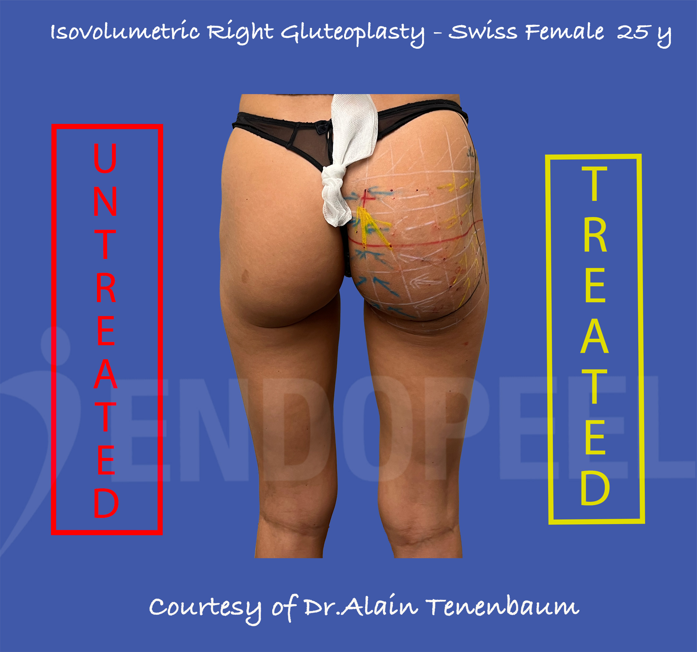
U01 · F03Result 1 h after treatment

Swiss female · right side treated

Swiss female unilateral gluteoplasty, patient right treated and left untreated
Original labels preserved: untreated left side and treated right side.
GREEN ARROWS

Internal widening

The green arrows in the internal column indicate the tensors for widening of the intergluteal cleft.

YELLOW ARROWS

External Latinisation

The yellow arrows in the two external columns indicate the tensors for external Latinisation.

LARGE YELLOW ARROW · 0.5–1 mL

Additional projection tensor

The large yellow arrow pointing toward the red cross indicates an additional tensor using 0.5–1 mL of the specific Endopeel® formulation, directed toward the point of maximal projection.
Isovolumetric reading · L × W × H
In this patient, lifting was combined with internal and external widening. Within the isovolumetric L × W × H framework, these contour changes would leave posterior projection lower than the intended normal projection without additional tensors directed toward the point of maximal projection. The additional tensor addresses this balance by reshaping the existing contour.
Indication
Projection and lower-contour refinement
Objective
Lifting, widening of the intergluteal cleft, external Latinisation and balanced maximal projection
Treated side
Patient’s right treated; left untreated
Sessions
1
Photographic interval
Result 1 h after treatment
Assessment
Standing posterior view
Interpretation limits
The untreated side is an internal comparator, not a randomized control. Pre-existing asymmetry, stance and muscular state can influence the apparent difference. The photograph does not establish equal baseline anatomy.
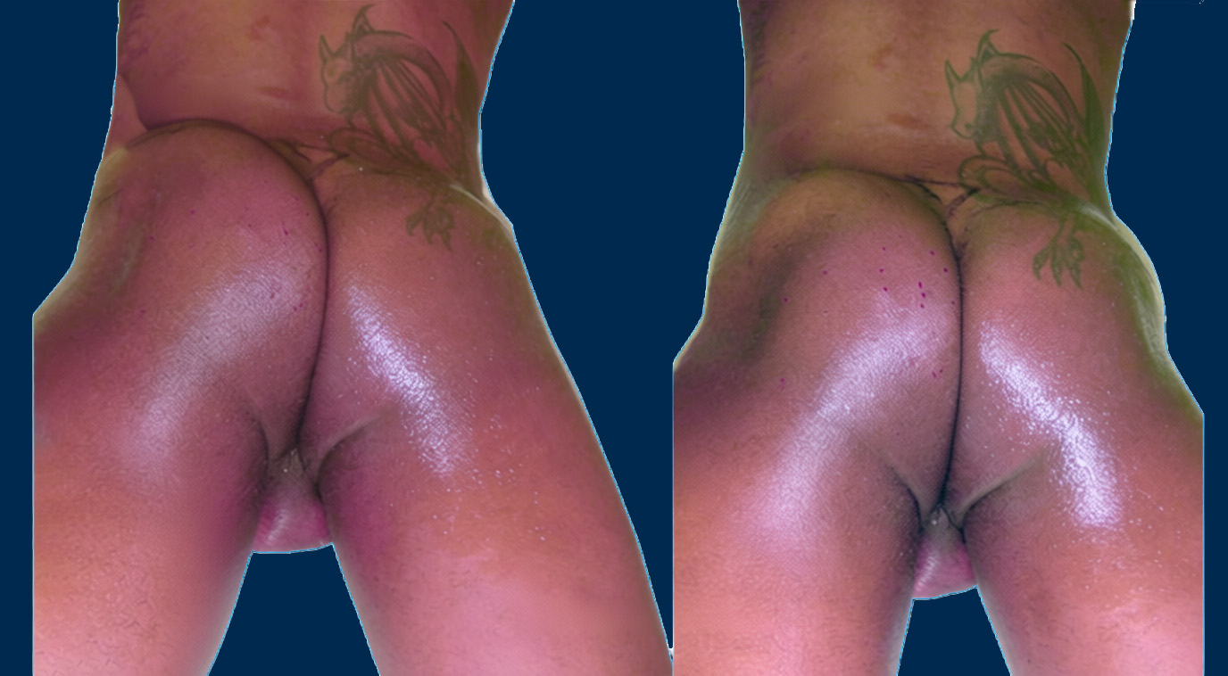
U02 · M01Result 1 h after treatment

Male dynamic contour · left side treated

Male unilateral gluteal contour during voluntary contraction, patient left treated and right untreated
Voluntary contraction · two complementary viewing angles.
Indication
Dynamic gluteal contour refinement
Objective
Assess side-specific contour under matched conditions
Treated side
Patient’s left treated; right untreated
Sessions
1
Photographic interval
Result 1 h after treatment
Assessment
Voluntary contraction · two complementary viewing angles
Interpretation limits
An immediate side-to-side observation does not prove the magnitude or durability of a treatment effect. Baseline asymmetry and positioning must be considered.

Clinical Results Atlas · 07 · Myomodulation®

Male & Athletic Gluteal Reshaping

The clinical objective follows the patient’s muscular development, proportions and preferred contour.
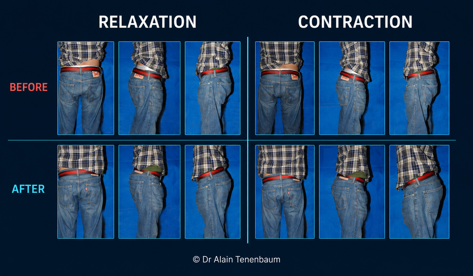
M02Result 1 h after treatment

Male contour in clothing · static and dynamic series

Male before-and-after gluteal results at relaxation and voluntary contraction
Original matrix: baseline and after treatment; posterior, oblique and profile views.
Indication
Male contour and projection refinement
Objective
Compare contour across viewing angles and muscular states
Treated side
Both
Sessions
1
Photographic interval
Result 1 h after treatment
Assessment
Posterior, oblique and profile · rest and contraction
Interpretation limits
The montage illustrates multiple views of one clinical series. It is not several independent patients. Clothing and muscle effort may influence apparent projection; no strength or performance outcome is measured.
ATHLETIC

Redistribution of an existing contour

In an already developed muscular contour, assess vertical compactness and the position of maximal projection while respecting width and overall proportions.

INDIVIDUAL

A defined objective for each patient

Athletic refinement, projection, symmetry and a softer contour are different objectives. A photograph alone cannot assign a patient’s fitness level or treatment goal.

Clinical Results Atlas · 08 · Myomodulation®

Dynamic Results — Rest vs Voluntary Contraction

The same contour can look different under different muscular conditions. Both states deserve a separate reading.
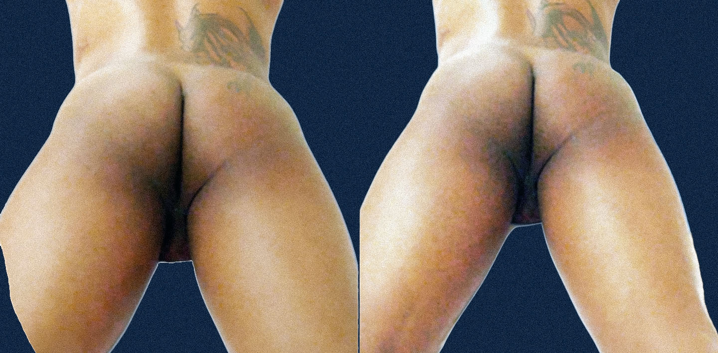
M01 · U02Result 1 h after treatment

Unilateral male comparison · two muscular states

Male gluteal contour at relaxation after unilateral treatment
RELAXATION · patient’s left treated; right untreated.
Male gluteal contour during voluntary contraction after unilateral treatment
CONTRACTION · patient’s left treated; right untreated.
Indication
Dynamic gluteal contour refinement
Objective
Read lateral contour and projection at rest and during contraction
Treated side
Patient’s left treated; right untreated
Sessions
1
Photographic interval
Result 1 h after treatment
Assessment
Rest and voluntary contraction · complementary angles
Interpretation limits
The photographic interval is confirmed by the clinical author. Contraction effort is not instrumentally measured; the photographs do not demonstrate increased muscular strength or long-term function.

Clinical Results Atlas · 09 · Myomodulation®

Asymmetry, Atrophy & Reconstructive Cases

A contour difference needs an anatomical explanation. Reconstructive observations must be interpreted within their individual clinical context.

R01Result 30 min after treatment

Post-traumatic gluteal atrophy · male case

Male post-traumatic gluteal atrophy before and 30 minutes after one Myomodulation session
Original clinical comparison · one session · 30-minute result.
Indication
Post-traumatic gluteal atrophy
Objective
Immediate visible contour correction
Treated side
Left
Sessions
1 session
Photographic interval
Result 30 min after treatment
Assessment
Original clinical comparison
Interpretation limits
An early change in contour does not demonstrate regeneration of muscle tissue, restoration of muscle mass or functional recovery. Trauma history, clinical examination and longitudinal review are necessary for reconstructive interpretation.
ASYMMETRY

Document both sides before treatment

Compare baseline height, width, projection and muscular response. A difference after unilateral treatment must not be mistaken automatically for a pre-existing defect.

RECONSTRUCTION

Keep contour and function distinct

Previous trauma, surgery or implants require case-specific assessment. These images support contour discussion; they do not establish functional recovery.

Clinical Results Atlas · 10 · Myomodulation®

Results in Clothing

Clothing reveals how gluteal projection interacts with everyday silhouette, pocket position and fabric tension.
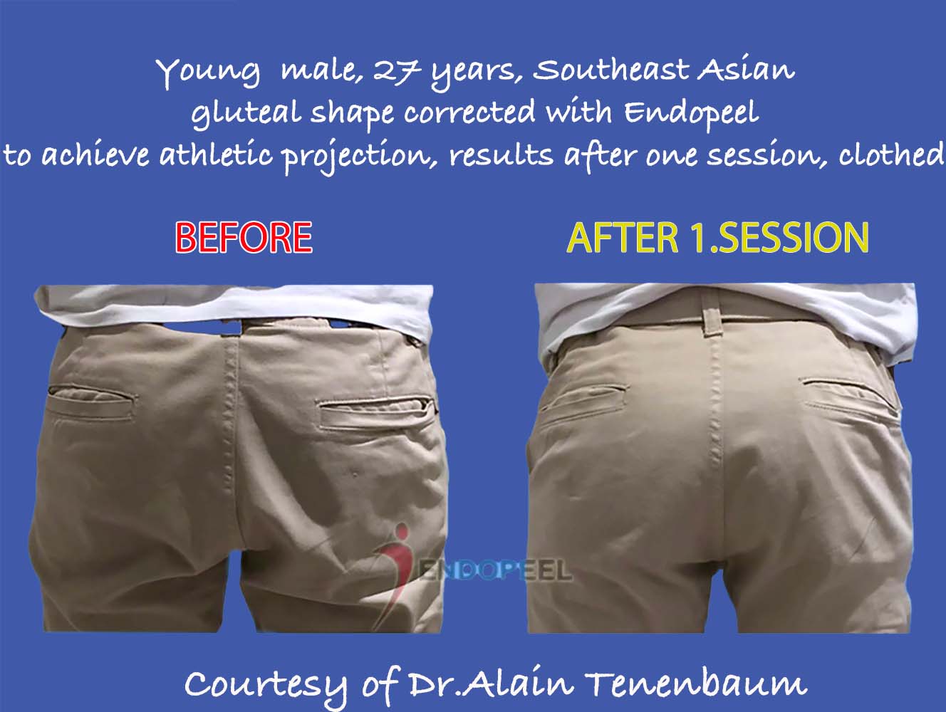
Male athletic gluteal projection in clothing before and after one session
Male athletic gluteal projection in clothing · before / after 1 session · Result 1 h after treatment.
POCKETS

An external reference

Pocket position is useful for visual comparison but is not a fixed anatomical landmark.

FABRIC

Match the garment

Fabric stretch, garment size, waistband height and posture can change the apparent lifting or projection.

ANATOMY

Pair clothed and nude views

When available, relate the garment silhouette to the actual gluteal landmarks and the treated side.

This male clinical comparison illustrates athletic gluteal projection in clothing after 1 session. Fabric tension, garment position and posture may influence the apparent change. Clothing comparisons alone cannot measure anatomical displacement.

Clinical Results Atlas · 11 · Myomodulation®

Immediate vs Follow-up Results

Time is part of the result. Immediate contour observations and later photographic comparisons answer different questions.

30 MIN

Post-traumatic atrophy

R01 documents one session and a 30-minute photograph.

1 H

Immediate case comparisons

The clinical author confirmed one hour for the other immediate cases in this atlas.

7 DAYS · 1 MONTH

Swiss photographic follow-up

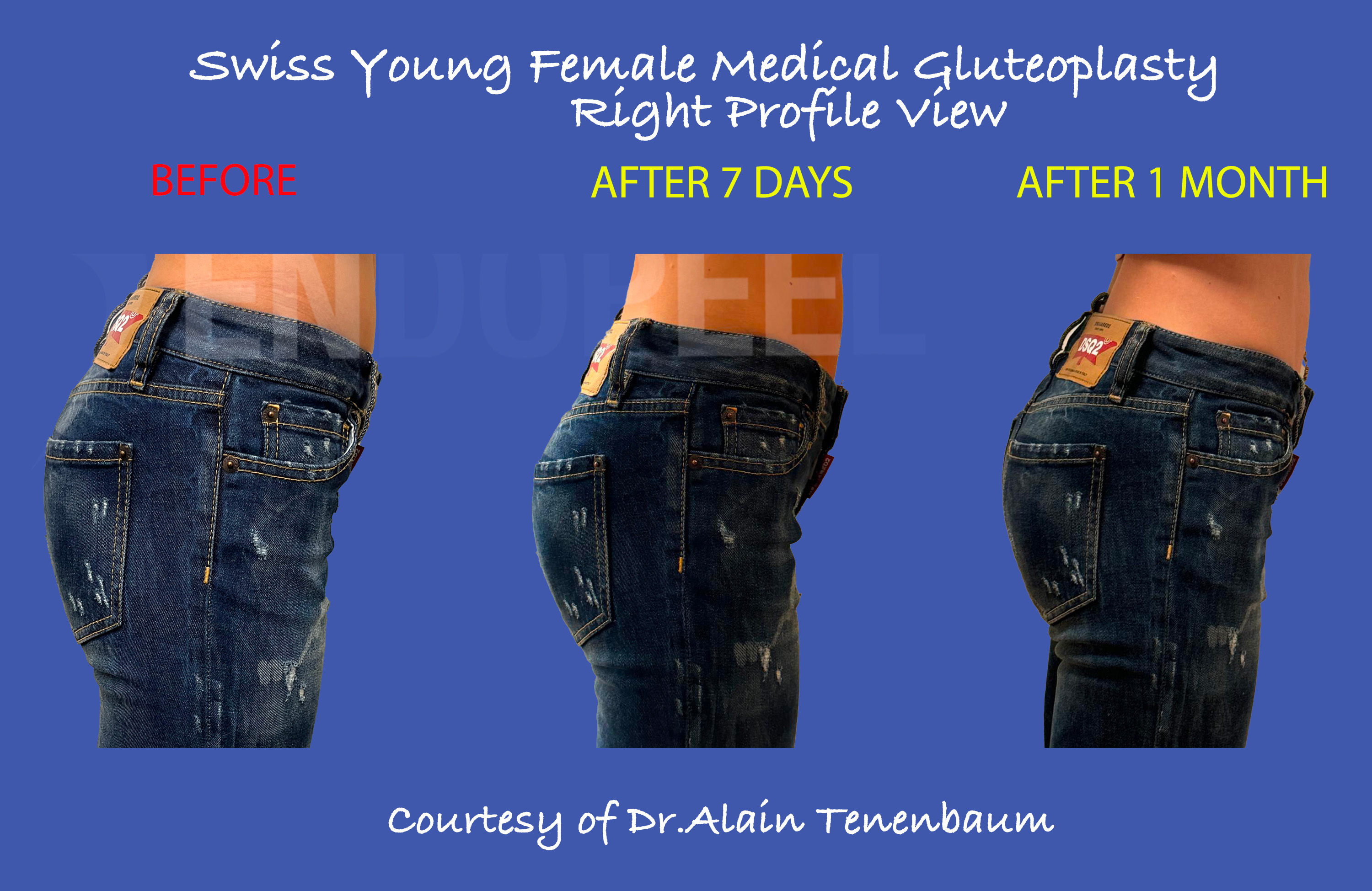
The selected archival series explicitly labels two later time points. These labels are retained exactly.
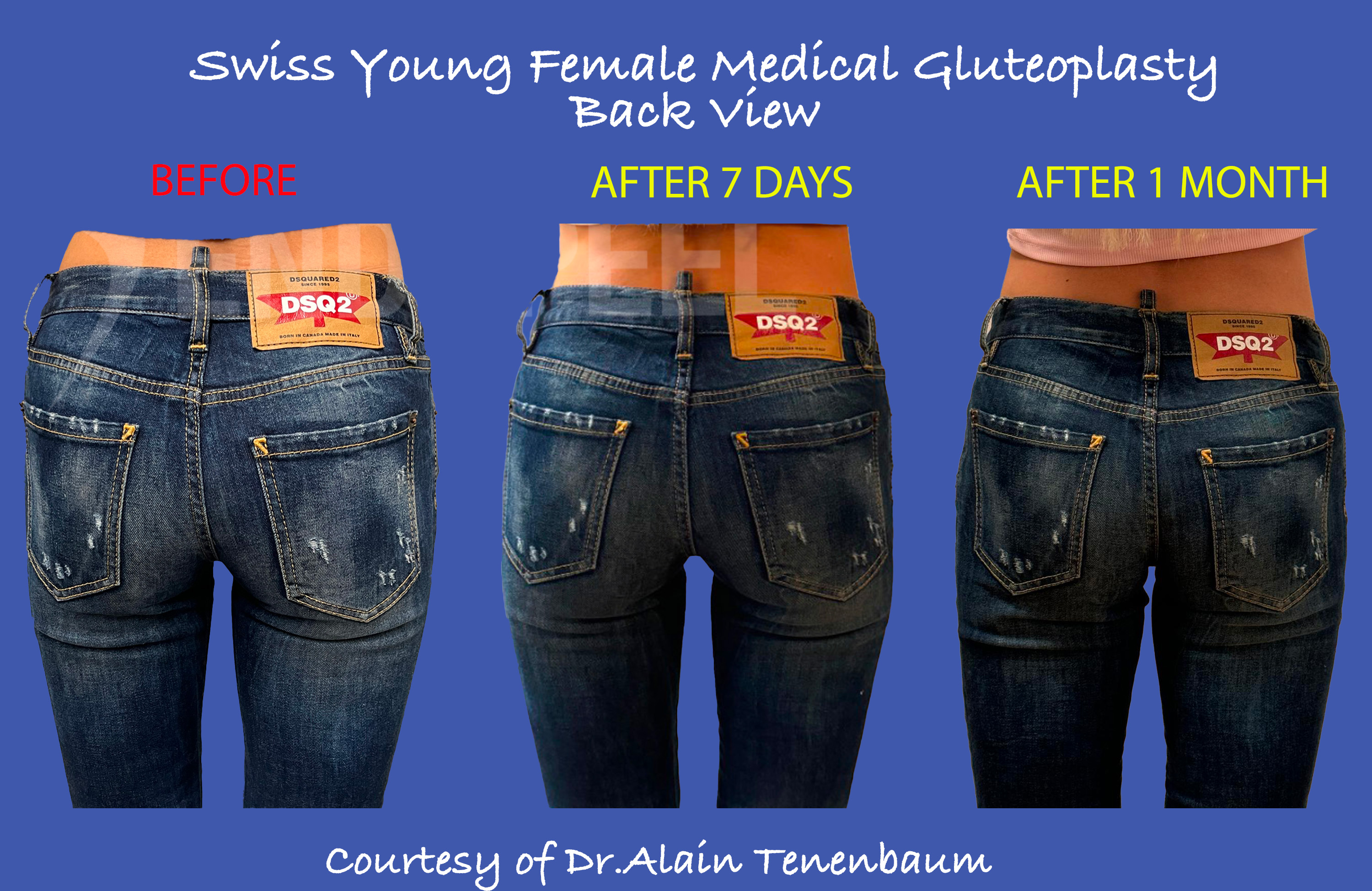
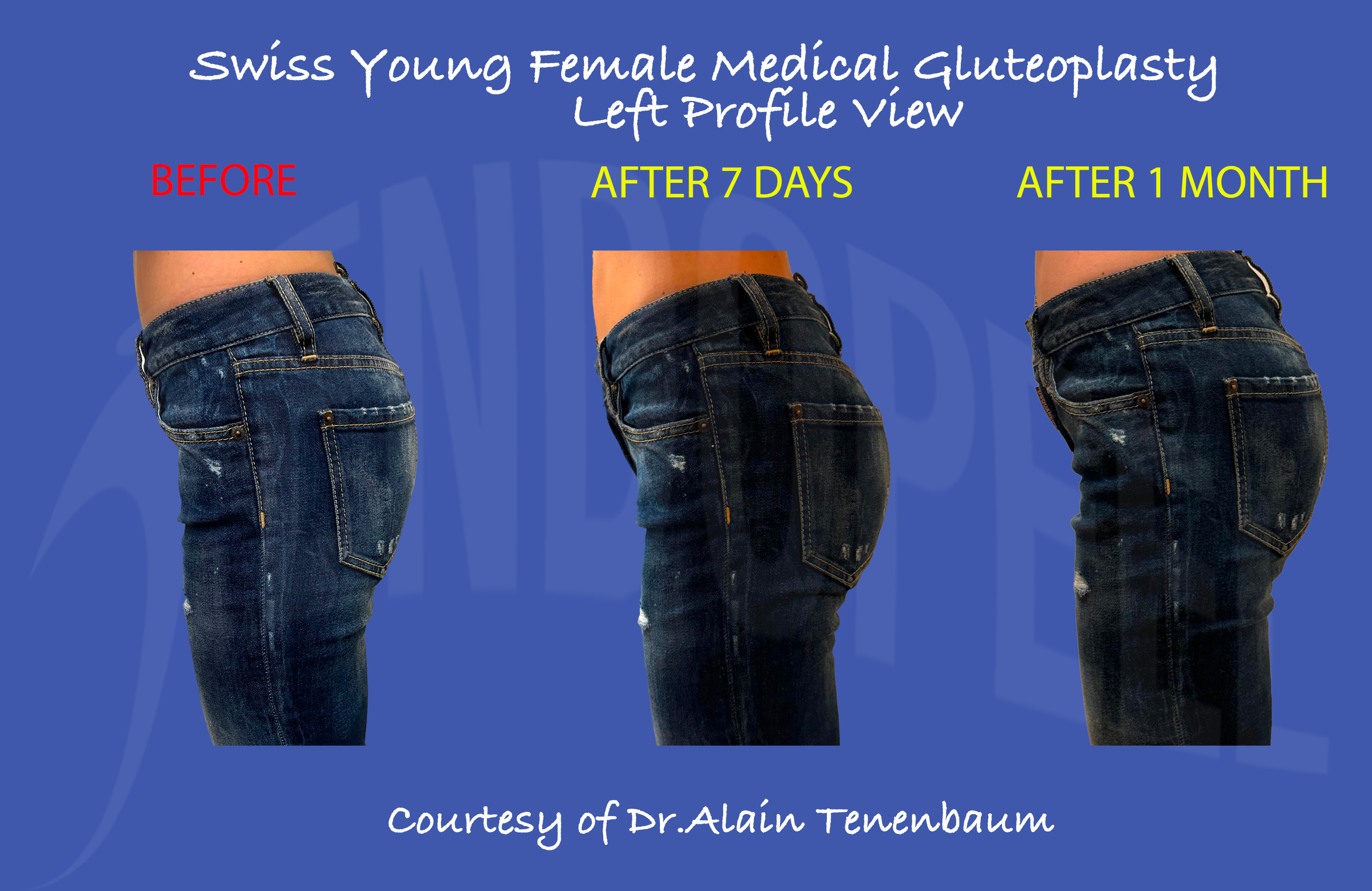
F03-FUBefore · 7 days · 1 month

Swiss female · archival follow-up series

Swiss female gluteal contour before treatment, after 7 days and after 1 month
Posterior series · BEFORE / AFTER 7 DAYS / AFTER 1 MONTH.
Swiss female left-profile contour before treatment, after 7 days and after 1 month
Left-profile series · BEFORE / AFTER 7 DAYS / AFTER 1 MONTH.
Swiss female right-profile contour before treatment, after 7 days and after 1 month
Right-profile series · BEFORE / AFTER 7 DAYS / AFTER 1 MONTH.
Indication
Female gluteal projection and contour follow-up
Objective
Compare evolution beyond the immediate photograph
Treated side
Both
Sessions
3
Photographic interval
Before · 7 days · 1 month
Assessment
Posterior and bilateral profile views
Interpretation limits
Intervals are taken from the labels embedded in these images. The series is presented separately from the 1-hour observations. Both sides were treated over 3 sessions. The timing of each session relative to the labelled photographs is not specified here. One month does not establish long-term durability.

Clinical Results Atlas · 12 · Myomodulation®

Clinical Interpretation & Limitations

Describe what can be observed, distinguish what is inferred, and reserve longitudinal conclusions for follow-up.

OBSERVE

Visible contour relationships

Projection, external contour, lower-fold position and side-to-side appearance can be described qualitatively.

STANDARDIZE

Photographic conditions

Match camera height, distance, focal length, lighting, pelvic position, garment and muscular state.

FOLLOW

Stability over time

An immediate photograph cannot establish maintenance needs, durability or a typical result across patients.

MEASURE

Volume and function separately

Exact volume conservation, muscle mass, strength and functional recovery require appropriate measurement beyond photographs.

CONTEXT

Keep the clinical history

Session count, previous procedures, baseline asymmetry and any complementary treatment affect interpretation.

INDIVIDUALIZE

Selected cases remain selected cases

This collection does not provide a denominator, complication rate or comparative efficacy estimate.

Clinical Results Atlas · 13 · Myomodulation®

Clinical Case Matrix

A compact clinical index. Repeated views and thematic re-use do not represent additional patients.

Case metadata and photographic context
CaseClinical indicationObjectiveTreated sideSessionsIntervalViews
F01Female projection & liftingProjection / lower contourLeft (profile)1 (posterior comparison)1 hProfile + posterior
F02Turkish female profileProfile projectionLeft11 hLeft profile
F03Swiss femaleProjection / contourBoth21 monthPosterior + left profile
U01Swiss female unilateral illustrationLifting / internal and external widening / projection balanceRight11 hStanding nude
F04Mexican female contourLatinisation / projection / lifting / intergluteal cleft and infragluteal diamond wideningBoth11 hClinical + clothing
M01 / U02Male dynamic contourRest / contractionLeft treated; right untreated11 hTwo angles / two states
M02Male clothed seriesProjection / dynamic contourBoth11 hPosterior / oblique / profile
R01Post-traumatic atrophyImmediate contour correctionLeft130 minOriginal comparison
F03-FUSwiss archival follow-upEvolution of clothed contourBoth37 days / 1 monthPosterior + profiles
“To be confirmed” identifies a field awaiting a case-specific confirmation. The atlas does not infer session count from the general treatment protocol. Intervals of 1 h and 30 min follow the clinical author’s confirmation; 7-day and 1-month intervals follow the archival image labels.

Clinical Results Atlas · 14 · Myomodulation®

Clinical Results FAQ

The clinical questions behind each photograph. Explore the contour, the treatment context and the time point with a precise, considered reading.

ISOVOLUMETRIC RESHAPINGCLINICAL INTERPRETATIONPHOTOGRAPHIC CONTEXT
01VOLUMEDoes greater projection mean added volume?

The isovolumetric objective is to reshape the existing contour. Greater posterior projection does not by itself indicate an increase in total volume. Read length, width and projection together through the L × W × H framework.

02MYOMODULATION®What is Endopeel® in this atlas?

Endopeel® is the specific formulation used for Myomodulation®. It is not a product: it is the formulation used within this approach to isovolumetric reshaping.

03PHOTOGRAPHIC INTERVALWhen were these results photographed?

R01 shows a result 30 minutes after treatment. The other immediate observations are presented at 1 hour. F03 is shown 1 month after treatment; the archival follow-up series in section 11 retains its labelled intervals of 7 days and 1 month.

04UNILATERAL COMPARISONWhy compare treated and untreated sides?

Comparing both sides in the same patient helps assess a side-specific contour at the same time point. Baseline asymmetry, stance and muscular state still affect interpretation. The untreated side provides an internal comparison rather than a randomized control.

05DYNAMIC CONTOURWhy show rest and voluntary contraction?

Muscular state changes the visible contour. Comparing rest with rest and contraction with contraction helps describe dynamic behaviour. A photographic difference does not measure muscle strength or functional recovery.

06TREATMENT HISTORYHow many sessions were performed?

F01 and F02 are shown after 1 session. F03 is shown after 2 sessions. F04, U01, M01/U02, M02 and R01 are shown after 1 session. The male clothed comparison in section 10 is labelled after 1 session, while the archival follow-up series in section 11 comprises 3 sessions.

07FOLLOW-UPDo these photographs prove long-term stability?

These photographs illustrate their stated time points. Immediate observations and a one-month comparison do not establish long-term stability. Longer follow-up under matched photographic conditions is needed to assess durability.

08CLOTHED RESULTSCan a clothed image establish anatomical lifting?

Clothing illustrates how the contour appears in everyday silhouette. Fabric tension, pocket placement, waistband height and posture influence the comparison. Corresponding anatomical views and clinical examination help interpret apparent lifting.

Clinical Results Atlas · 15 · Myomodulation®

Continue the Clinical Journey

Connect the atlas to anatomical principles, the technical sequence and professional training.

SCIENCE

Understand the framework

Revisit length, width, projection and the position of maximal projection.

TECHNIQUE

Follow the clinical sequence

Explore anatomical landmarks, individualized mapping and the step-by-step approach.

TRAINING

Discuss cases in a workshop

Relate image interpretation to assessment and practical clinical planning.